. 2002 Apr 15;157(2):211-7.
doi: 10.1083/jcb.200201098. Epub 2002 Apr 15.

An inactive pool of GSK-3 at the leading edge of growth cones is implicated in Semaphorin 3A signaling

Affiliations

An inactive pool of GSK-3 at the leading edge of growth cones is implicated in Semaphorin 3A signaling

Britta J Eickholt et al. J Cell Biol. .

Abstract

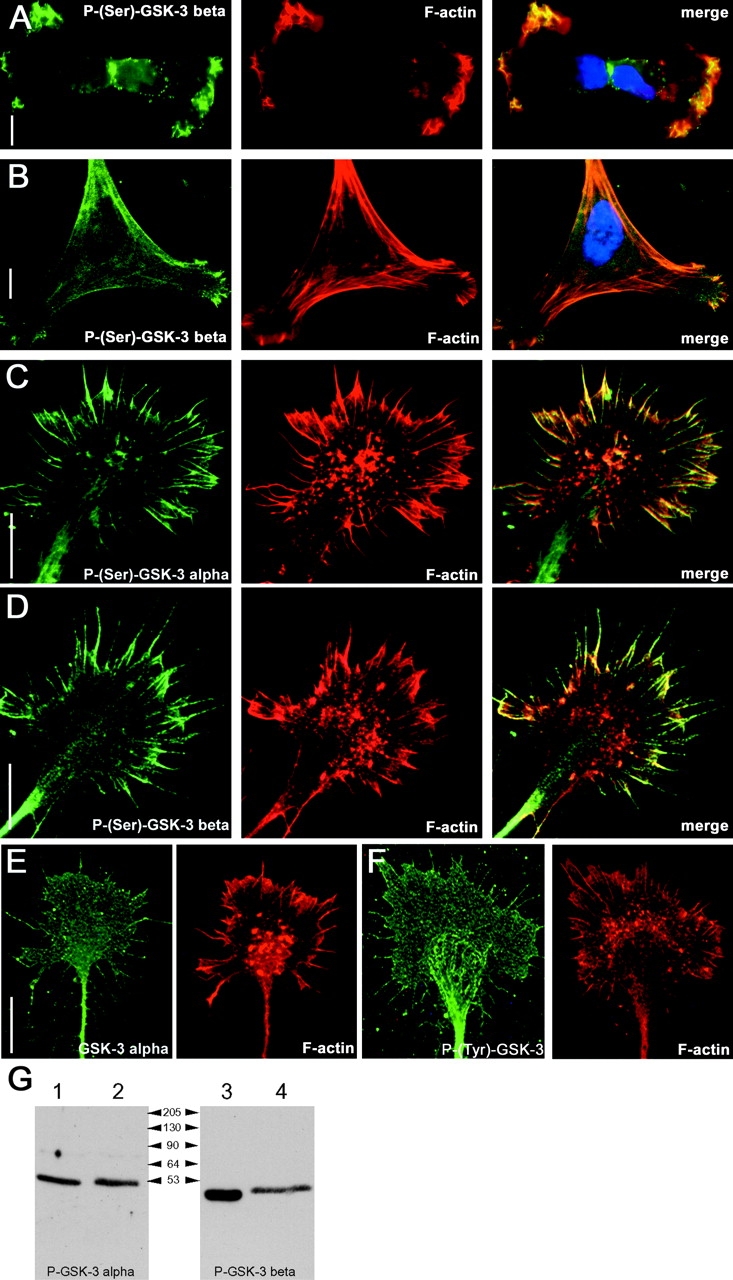
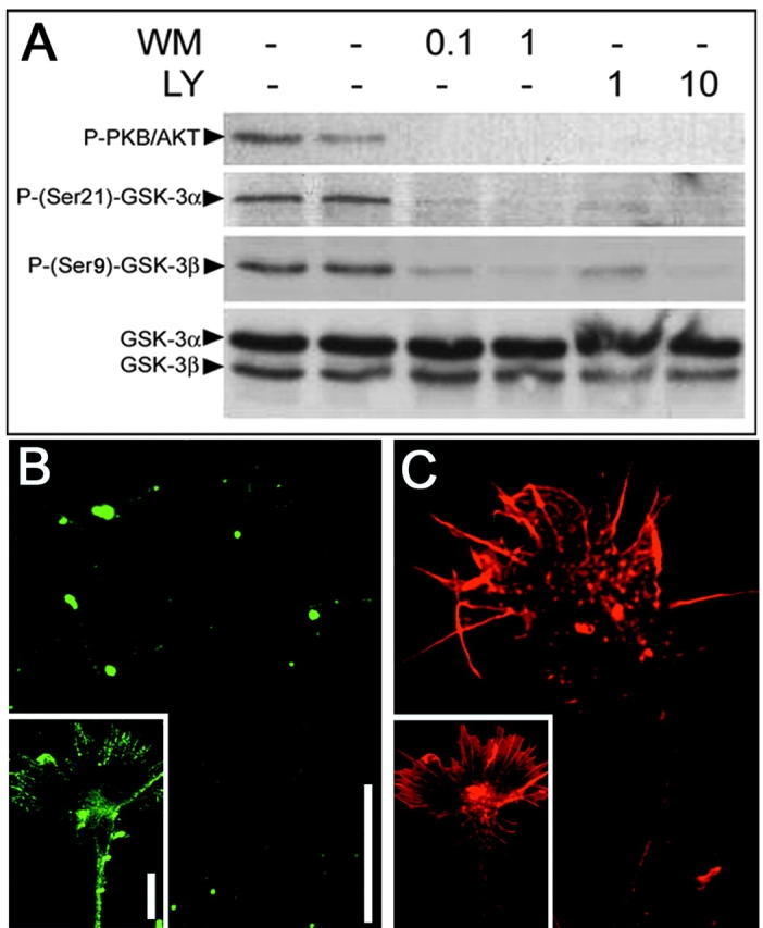
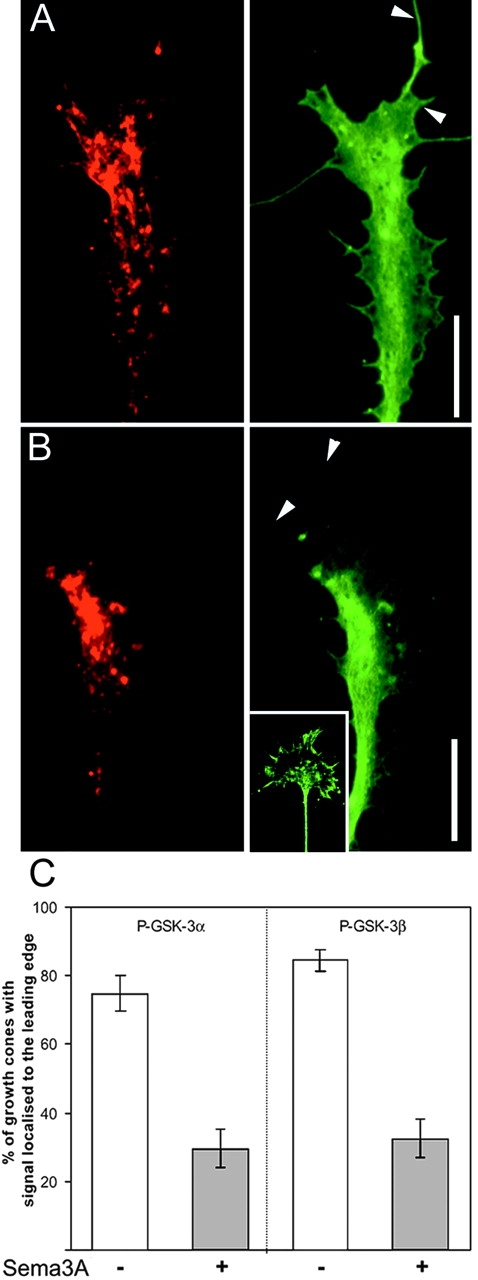
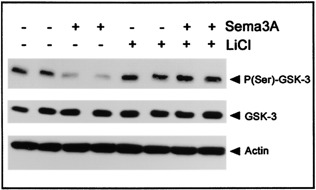
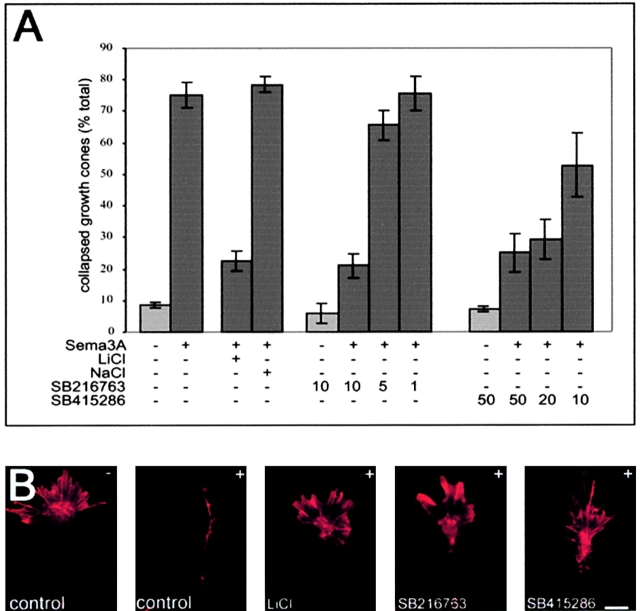
Glycogen synthase kinase (GSK)-3 is a serine/threonine kinase that has been implicated in several aspects in embryonic development and several growth factor signaling cascades. We now report that an inactive phosphorylated pool of the enzyme colocalizes with F-actin in both neuronal and nonneuronal cells. Semaphorin 3A (Sema 3A), a molecule that inhibits axonal growth, activates GSK-3 at the leading edge of neuronal growth cones and in Sema 3A-responsive human breast cancer cells, suggesting that GSK-3 activity might play a role in coupling Sema 3A signaling to changes in cell motility. We show that three different GSK-3 antagonists (LiCl, SB-216763, and SB-415286) can inhibit the growth cone collapse response induced by Sema 3A. These studies reveal a novel compartmentalization of inactive GSK-3 in cells and demonstrate for the first time a requirement for GSK-3 activity in the Sema 3A signal transduction pathway.


Figures

Figure 1.
Figure 1.
GSK-3 is maintained inactive at the leading edge of migratory cells and colocalizes with F-actin. Distribution of P-(Ser9)-GSK-3β immunoreactivity in MDA-MB-231 breast carcinoma cells (A, left) and in primary chick fibroblast (B, left). A parallel-performed phalloidin staining (middle) reveals a great overlap of inactive serine-phosphorylated GSK-3 with F-actin (right, merge). Likewise, in DRG growth cones the signals detected using an anti–P-(Ser21)-GSK-3α antibody (C) and an anti–P-(Ser9)-GSK-3β (D) are found colocalized with F-actin in the filopodia and at the leading edge of the lamellipodia. Stainings performed using a pan GSK-3α (E) and a P-(Y)-GSK-3 (F) antibody reveal that GSK-3 in present throughout the entire growth cone structure. Bars, 15 μm. (G) Western blots probed with indicated P-(Ser)–specific antibodies: lanes 1 and 3 show chick brain lysate, and lanes 2 and 4 show Cos-7 cell lysate that have been transfected with GSK-3α and GSK-3β, respectively.
Figure 2.
Figure 2.
Dephosphosphorylation of GSK-3 by inhibition of PI 3-kinase. (A) In primary DRG neurons, treatment with wortmannin (WM) or LY294002 (LY) at given μM concentrations for 1 h reduces the phosphorylation of PKB/AKT and both GSK-3α and GSK-3β. (B) Treatment of DRG explant with wortmannin at 0.1 μM results in a loss of the P-Ser(9)-GSK-3β signal seen in untreated control cultures (insert). (C) In a parallel-performed phalloidin staining, the growth cone is clearly visible. Bars, 15 μm.
Figure 3.
Figure 3.
Sema 3A activates GSK-3 at the leading edge of sensory growth cones. DRG cultures grown overnight were treated with a Sema 3A–Fc chimera for 2 min before fixation. Growth cones were stained with phalloidin–Texas red (left) and anti–GSK-3α (A, right) and with phalloidin–Texas red (left) and anti–P-(Ser21)-GSK-3α (B, right). After exposure to Sema 3A, the entire GSK-3α pool remains homogeneously distributed throughout the growth cone and is still detectable in filopodia, whereas the inactive P-(Ser21)-GSK-3α is depleted at the leading edge of the growth cone and filopodia (arrowheads). The insert shows the parallel performed P-(Ser21)-GSK-3α staining in untreated control cultures. Bars, 15 μm. (C) Percentage of growth cones showing enriched staining at the leading edge before and after treatment with Sema 3A–Fc. Bars show ± SEM.
Figure 4.
Figure 4.
Decrease in phosphorylation of GSK-3 after Sema 3A treatment in MDA-MB-231 breast carcinoma cell lines. Western blots of MDA-MB 231 cell extracts using anti–P-(Ser9)-GSK-3β, anti–GSK-3β, and antiactin antibodies. Cell lysates were prepared from PBS-treated control and Sema 3A–Fc–treated (1 μg/ml for 1 h) MDA-MB 231 cells in the presence or absence of LiCl (at 20 mM). All experiments were performed in duplicates. Sema 3A treatment notably decreases Ser9 phosphorylation of GSK-3. Treatment with the GSK-3 inhibitor LiCl enhances GSK-3 phosphorylation slightly and antagonizes the Sema 3A–mediated decrease in Ser9 phosphorylation. Blots were reprobed with an anti–GSK-3β antibody to confirm equal loading of samples.
Figure 5.
Figure 5.
Inhibition of GSK-3 prevents Sema 3A–induced growth cone collapse. (A) Addition of Sema 3A–Fc to DRG explant cultures for 30 min induces a growth collapse response. The graphs show the percentage of collapsed growth cone ± SEM (n ≥ 4 independent experiments). In each experiment ≥100 growth cones were counted. In the presence of 20 mM LiCl, the Sema 3A response is substantially inhibited, whereas NaCl at 20 mM did not alter the Sema 3A–induced growth cone collapse. Likewise, the two specific GSK-3 inhibitors, SB-216763 and SB-415286 (used at μM concentrations as stated), inhibited the Sema 3A–induced growth cone collapse in a dose-dependent manner. (B) Examples of phalloidin-stained DRG growth cones in order to visualize the distribution of F-actin. First micrograph shows a control untreated (−) growth cone; all subsequent pictures show growth cones that have been treated with Sema 3A (+) in the absence (control) and presence of LiCl (20 mM), SB-216763 (10 μM), and SB-415286 (30 μM). In the presence of the GSK-3 antagonist, growth cone collapse is clearly inhibited. Bar, 15 μm.

References

    1. Aizawa, H., S. Wakatsuki, A. Ishii, K. Moriyama, Y. Sasaki, K. Ohashi, Y. Sekine-Aizawa, A. Sehara-Fujisawa, K. Mizuno, Y. Goshima, et al. 2001. Phosphorylation of cofilin by LIM-kinase is necessary for semaphorin 3A-induced growth cone collapse. Nat. Neurosci. 4:367–373. - PubMed
    1. Bagnard, D., C. Vaillant, S.T. Khuth, N. Dufay, M. Lohrum, A.W. Puschel, M.F. Belin, J. Bolz, and N. Thomasset. 2001. Semaphorin 3A-vascular endothelial growth factor-165 balance mediates migration and apoptosis of neural progenitor cells by the recruitment of shared receptor. J. Neurosci. 21:3332–3341. - PMC - PubMed
    1. Baum, L., R. Seger, J.R. Woodgett, S. Kawabata, K. Maruyama, M. Koyama, J. Silver, and T. Saitoh. 1995. Overexpressed tau protein in cultured cells is phosphorylated without formation of PHF: implication of phosphoprotein phosphatase involvement. Brain Res. Mol. Brain Res. 34:1–17. - PubMed
    1. Coghlan, M.P., A.A. Culbert, D.A. Cross, S.L. Corcoran, J.W. Yates, N.J. Pearce, O.L. Rausch, G.J. Murphy, P.S. Carter, L. Roxbee Cox, et al. 2000. Selective small molecule inhibitors of glycogen synthase kinase-3 modulate glycogen metabolism and gene transcription. Chem. Biol. 7:793–803. - PubMed
    1. Cook, D., M.J. Fry, K. Hughes, R. Sumathipala, J.R. Woodgett, and T.C. Dale. 1996. Wingless inactivates glycogen synthase kinase-3 via an intracellular signalling pathway which involves a protein kinase C. EMBO J. 15:4526–4536. - PMC - PubMed

Publication types

MeSH terms