. 2002 Apr;109(8):1057-63.
doi: 10.1172/JCI14211.

Increased vascular permeability in C1 inhibitor-deficient mice mediated by the bradykinin type 2 receptor

Affiliations

Increased vascular permeability in C1 inhibitor-deficient mice mediated by the bradykinin type 2 receptor

Eun D Han et al. J Clin Invest. 2002 Apr.

Abstract

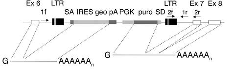
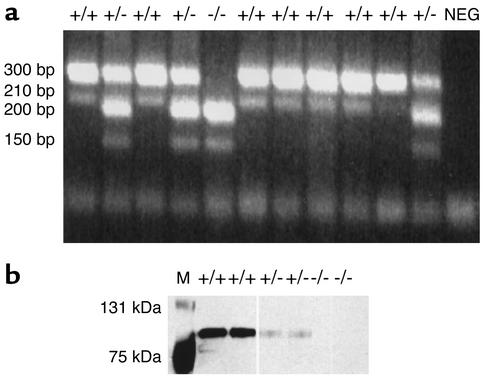
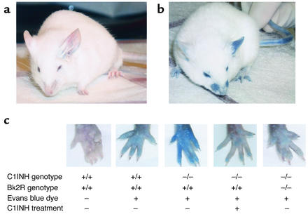
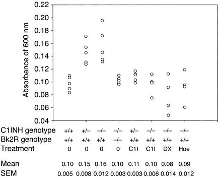
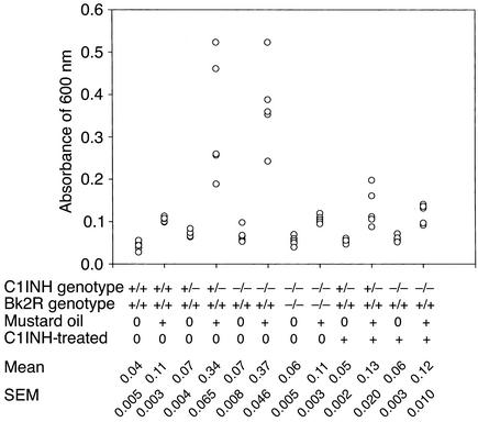
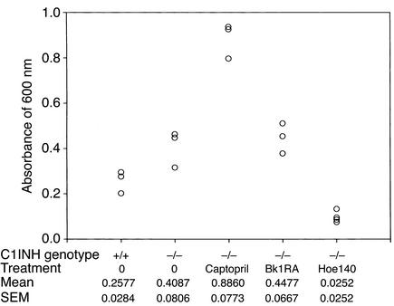
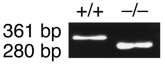
Heterozygosity for C1 inhibitor (C1INH) deficiency results in hereditary angioedema. Disruption of the C1INH gene by gene trapping enabled the generation of homozygous- and heterozygous-deficient mice. Mating of heterozygous-deficient mice resulted in the expected 1:2:1 ratio of wild-type, heterozygous, and homozygous-deficient offspring. C1INH-deficient mice showed no obvious phenotypic abnormality. However, following injection with Evans blue dye, both homozygous and heterozygous C1INH-deficient mice revealed increased vascular permeability in comparison with wild-type littermates. This increased vascular permeability was reversed by treatment with intravenous human C1INH, with a Kunitz domain plasma kallikrein inhibitor (DX88), and with a bradykinin type 2 receptor (Bk2R) antagonist (Hoe140). In addition, treatment of the C1INH-deficient mice with an angiotensin-converting enzyme inhibitor (captopril) increased the vascular permeability. Mice with deficiency of both C1INH and Bk2R demonstrated diminished vascular permeability in comparison with C1INH-deficient, Bk2R-sufficient mice. These data support the hypothesis that angioedema is mediated by bradykinin via Bk2R.


Figures

Figure 1
Figure 1
Targeting the C1INH gene in mice by random insertional mutagenesis (12). The white boxes indicate C1INH gene exons (Ex). The retroviral vector with the galactosidase/neomycin phosphotransferase fusion gene (geo), the puromycin N-acetyl transferase gene (puro), and other components are inserted within intron 6, 210 bp in the 5′ direction from exon 7, and are indicated with shaded boxes. 1f, 2f, 1r, and 2r represent PCR primers. LTR, long term repeat; SA, splice acceptor sequence; IRES, internal ribosome entry site; pA, polyadenylation sequence; PGK, phosphoglycerate kinase promoter; SD, splice donor sequence.
Figure 2
Figure 2
Genotype/phenotype analysis. (a) The genotype of the C1INH-targeted mice was determined by PCR amplification using the two primer pairs described in Methods. Amplification in the C1INH+/+ mice resulted in bands of 300 bp and 210 bp; in the C1INH+/– mice, bands of 300 bp, 210 bp, 200 bp, and 150 bp resulted; in the C1INH–/– mice, bands of 200 bp and 150 bp resulted. (b) Detection of C1INH by Western blot analysis of mouse serum using antiserum to mouse C1INH. NEG, negative control; M, molecular weight marker.
Figure 3
Figure 3
Analysis of vascular permeability. Extravasation of Evans blue dye at 15–30 minutes was much more extensive in C1INH-deficient mice (b) than in wild-type mice (a), particularly after the application of mustard oil to the left ears of the mice. (c) The difference in the amount of extravasation was clearly demonstrated by the rear footpads of mice of each genotype. Administration of human C1INH resulted in reduced vascular permeability, as did the combination of Bk2R deficiency together with C1INH deficiency.
Figure 4
Figure 4
Spectrophotometric analysis of vascular permeability in footpads. Quantitation of Evans blue dye extracted from the paws of mice of the indicated genotypes, either treated or not treated with intravenous C1INH (C1I), DX88 (DX), or Hoe140 (Hoe).
Figure 5
Figure 5
Spectrophotometric analysis of the vascular permeability response to application of mustard oil to the ears of mice of the indicated phenotypes and treatments.
Figure 6
Figure 6
Spectrophotometric analysis of vascular permeability in the small intestine. Quantitation of extravasated Evans blue dye in C1INH–/– mice treated with an ACE inhibitor (captopril), a Bk1R antagonist (des-Arg9,[Leu8]-bradykinin; Bk1RA), and a Bk2R antagonist (Hoe140).
Figure 7
Figure 7
Genotype analysis of C1INH–/–, Bk2R–/– mice. The genotypes of the double knockout mice were determined by PCR amplification using the primer pairs indicated in Methods. Amplification in the C1INH+/+, Bk2R+/+ mice resulted in a C1INH+/+ genotype as shown in Figure 2a, while the Bk2R+/+ genotype showed a single band of 361 bp. Amplification in the C1INH–/–, Bk2R–/– mice resulted in a C1INH–/– genotype as shown in Figure 2a, while the Bk2R–/– genotype showed a single band of 280 bp.

Comment in

References

    1. Sim RB, Reboul A, Arlaud GJ, Villiers CL, Colomb MG. Interaction of 125I-labeled complement components C1r and C1s with protease inhibitors in plasma. FEBS Lett. 1979;97:111–115. - PubMed
    1. Ratnoff O, Pensky J, Ogston D, Naff G. The inhibition of plasmin, plasma kallikrein, plasma permeability factor, and the C’1r subcomponent of complement by serum C’1 esterase inhibitor. J Exp Med. 1969;129:315–331. - PMC - PubMed
    1. Forbes C, Pensky J, Ratnoff O. Inhibition of activated Hageman factor and activated plasma thromboplastin antecedent by purified C1 inactivator. J Lab Clin Med. 1970;76:809–815. - PubMed
    1. Gigli I, Mason JW, Colman RW, Austen KF. Interaction of plasma kallikrein with the C1 inhibitor. J Immunol. 1970;104:574–581. - PubMed
    1. Schreiber AD, Kaplan AP, Austen KF. Inhibition by C1INH of Hageman factor fragment activation of coagulation, fibrinolysis, and kinin generation. J Clin Invest. 1973;52:1402–1409. - PMC - PubMed

Publication types

MeSH terms