. 2002 Apr 15;195(8):1013-21.
doi: 10.1084/jem.20011963.

Presentation of the same glycolipid by different CD1 molecules

Affiliations

Presentation of the same glycolipid by different CD1 molecules

A Shamshiev et al. J Exp Med. .

Abstract

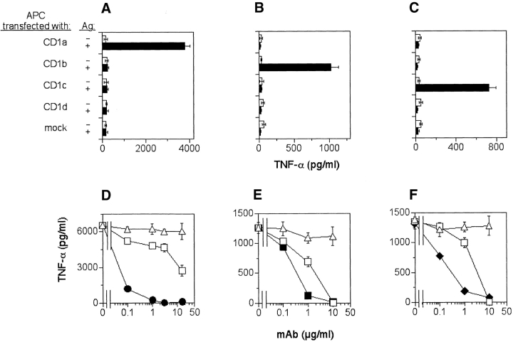
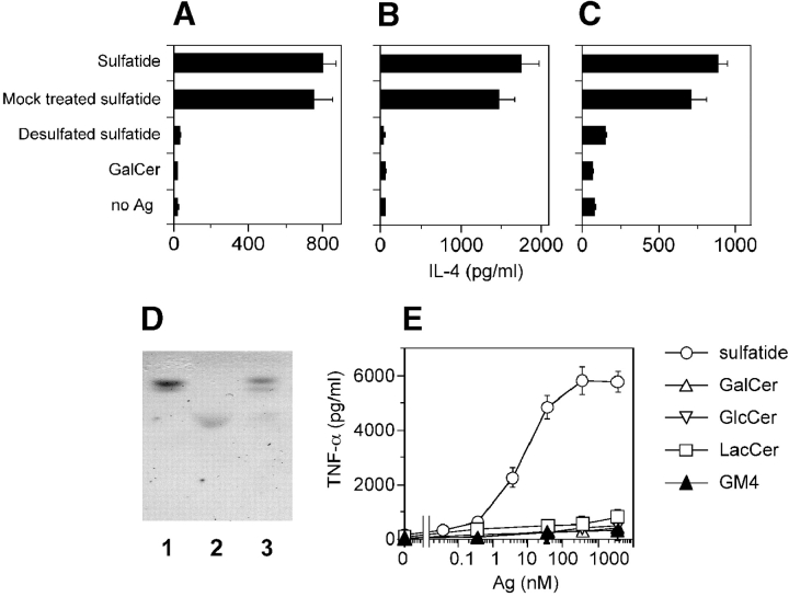
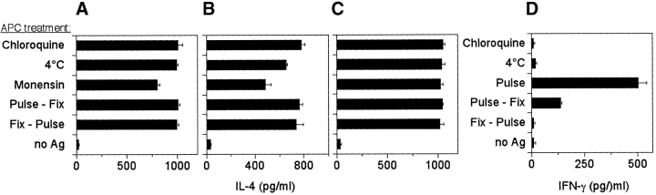
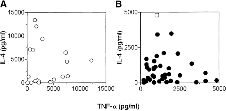
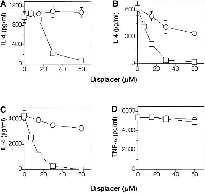
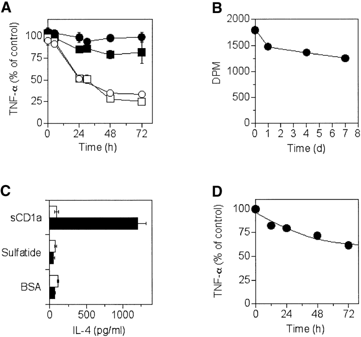
Five CD1 molecules are expressed in humans and it is unclear whether they have specialized or redundant functions. We found that sulfatide is a promiscuous CD1-binding ligand and have isolated T cell clones that are specific for sulfatide and restricted by distinct CD1 molecules. These clones have been used to compare the capacity of different CD1 to present the same glycolipid, to induce effector functions, and to form persistent immunogenic complexes. CD1a, CD1b, and CD1c molecules similarly load sulfatide on the cell surface without processing, and prime Th1 and Th2 responses. Stimulation by sulfatide-loaded CD1a persists much longer than that by CD1b and CD1c in living cells. Use of recombinant soluble CD1a confirmed the prolonged capacity to stimulate T cells. Moreover, other glycosphingolipids bind to all CD1, which suggests the presence of additional promiscuous ligands. Thus, group I CD1 molecules present an overlapping set of self-glycolipids, even though they are quite divergent from an evolutionary point of view.


Figures

Figure 1.
Figure 1.
Presentation of sulfatide by CD1a, CD1b, and CD1c antigen-presenting molecules. (A–C) CD1a-, CD1b-, CD1c-, CD1d-, or mock-transfected C1R cells were pulsed with 10 μM of sulfatide and used to stimulate the following T cell clones: K34B9.1 (A and D, CD1a restricted), DS1C9b (B and E, CD1b restricted) and DS1B9c (C and F, CD1c restricted). Stimulation in the presence (solid bars) or the absence (open bars) of sulfatide is shown. (D–F) Presentation of sulfatide by DC to the specific T cell clones was blocked by anti-CD1a (•), anti-CD1b (▪), anti-CD1c (♦), or anti-TCR Fab (□) fragments, but not by isotype-matched control mAbs (▵).
Figure 2.
Figure 2.
Fine antigen specificity of sulfatide-specific T cell clones. (A–C) Desulfation abolishes immunogenicity of sulfatide. DC were incubated with 10 μM of sulfatide, mock-treated sulfatide, desulfated sulfatide, or GalCer before the addition of the T cell clones K34B9.1 (A, CD1a restricted), DS1C9b (B, CD1b restricted), and DS1B9c (C, CD1c restricted). (D) Thin layer chromatographic analysis of GalCer (lane 1), sulfatide (lane 2), and sulfatide after desulfation (lane 3). (E) Dose response of a representative T cell clone (K34B9.1, CD1a restricted) stimulated with DC pulsed with sulfatide, GalCer, GlcCer, LacCer, and ganglioside GM4. Similar results were obtained with most of the clones and also with IL-4 ELISA (unpublished data). The results are representative of three independent experiments.
Figure 3.
Figure 3.
CD1a, CD1b, and CD1c molecules do not require internalization and endosomal acidification for sulfatide presentation. DC were pretreated under different conditions (as described in Materials and Methods), and then pulsed with sulfatide and fixed before the addition of the T cell clones K34B9.1 (A, CD1a restricted), DS1C9b (B, CD1b restricted), and DS1B9c (C, CD1c restricted). (D) The same treatments completely abolished the response of the clone GP2.7, which is specific for PPD and HLA-DR restricted. Fix-Pulse stands for the fixation of APC before pulsing with the antigen. Results are representative of three independent experiments.
Figure 4.
Figure 4.
Functional phenotype of CD1-restricted and sulfatide-specific T cell clones. DC pulsed with 10 μM of sulfatide were used to stimulate (A) CD1a- (○), (B) CD1b- (•), or CD1c-restricted (□) T cell clones. After 36 h the released IL-4 and TNF-α were detected by ELISA. Results from 58 clones are shown.
Figure 5.
Figure 5.
Sulfatide previously bound to CD1 is displaced by other glycolipids. (A–C) Fixed DC pulsed with high doses of sulfatide (60 μM) and then incubated with various doses of GM1 (□) or sphingomyelin (○) were used to stimulate the T cell clones K34B9.1 (A, CD1a restricted), DS1C9b (B, CD1b restricted), and DS1B9c (C, CD1c restricted). (D) To demonstrate the lack of toxicity, control Vγ9Vδ2 T cells activated by isopentenyl pyrophosphate were included. The data shown is representative of three independent experiments.
Figure 6.
Figure 6.
Persistence and stability of CD1–sulfatide complexes. (A) DC pulsed with 60 μM of sulfatide were used to stimulate the CD1a-restricted T cell clones K34B9.1 and DS1A16a (• and ▪, respectively), the CD1b-restricted clone DS1C9b (□), or the CD1c-restricted clone DS1B9c (○). Results are expressed as a percentage of controls (as described in Materials and Methods). (B) Soluble CD1a–[14C]sulfatide complexes were incubated for the indicated time at room temperature and the remaining radioactivity associated with sCD1a was measured after size exclusion chromatography (as described in Materials and Methods). (C) Soluble CD1a preincubated with sulfatide was immobilized and used to stimulate the CD1a-restricted sulfatide-specific T cell clone K34B9.1 (solid bars) or the MHC class II–restricted PPD-specific GP2.9 clone (open bars). Immobilized BSA or sulfatide were used as negative controls. (D) Immobilized sCD1a–sulfatide complexes were chased for the indicated time and used to stimulate the CD1a-restricted T cell clone K34B9.1 (as described in Materials and Methods).

References

    1. Porcelli, S.A., and R.L. Modlin. 1999. The CD1 system: antigen-presenting molecules for T cell recognition of lipids and glycolipids. Annu. Rev. Immunol. 17:297–329. - PubMed
    1. Park, S.H., and A. Bendelac. 2000. CD1-restricted T-cell responses and microbial infection. Nature. 406:788–792. - PubMed
    1. Matsuda, J.L., and M. Kronenberg. 2001. Presentation of self and microbial lipids by CD1 molecules. Curr. Opin. Immunol. 13:19–25. - PubMed
    1. Sieling, P.A., D. Chatterjee, S.A. Porcelli, T.I. Prigozy, R.J. Mazzaccaro, T. Soriano, B.R. Bloom, M.B. Brenner, M. Kronenberg, P.J. Brennan, et al. 1995. CD1-restricted T cell recognition of microbial lipoglycan antigens. Science. 269:227–230. - PubMed
    1. Burdin, N., L. Brossay, Y. Koezuka, S.T. Smiley, M.J. Grusby, M. Gui, M. Taniguchi, K. Hayakawa, and M. Kronenberg. 1998. Selective ability of mouse CD1 to present glycolipids: alpha-galactosylceramide specifically stimulates V alpha 14+ NK T lymphocytes. J. Immunol. 161:3271–3281. - PubMed

Publication types