. 2002 Apr 15;16(8):919-32.
doi: 10.1101/gad.973302.

SETDB1: a novel KAP-1-associated histone H3, lysine 9-specific methyltransferase that contributes to HP1-mediated silencing of euchromatic genes by KRAB zinc-finger proteins

Affiliations

SETDB1: a novel KAP-1-associated histone H3, lysine 9-specific methyltransferase that contributes to HP1-mediated silencing of euchromatic genes by KRAB zinc-finger proteins

David C Schultz et al. Genes Dev. .

Abstract

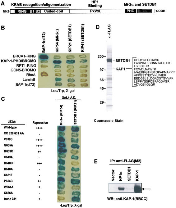
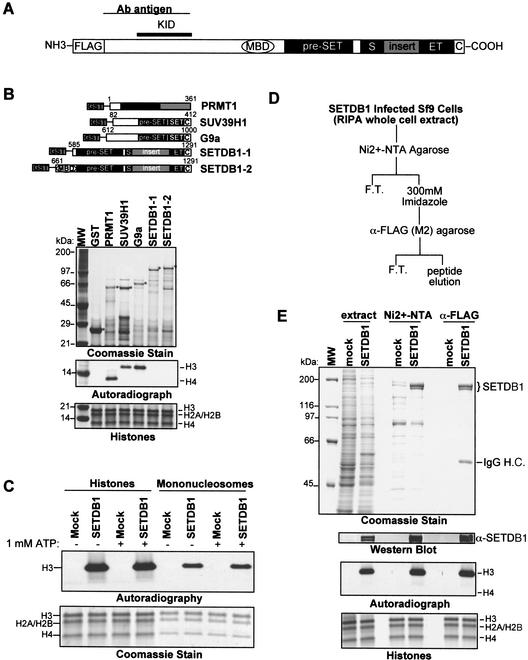
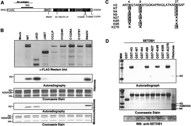
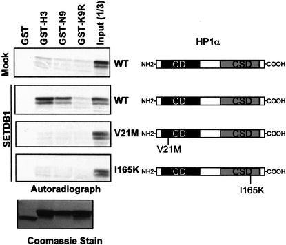
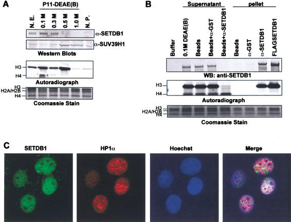
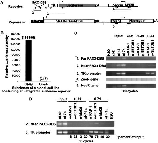
Posttranslational modification of histones has emerged as a key regulatory signal in eukaryotic gene expression. Recent genetic and biochemical studies link H3-lysine 9 (H3-K9) methylation to HP1-mediated heterochromatin formation and gene silencing. However, the mechanisms that target and coordinate these activities to specific genes is poorly understood. Here we report that the KAP-1 corepressor for the KRAB-ZFP superfamily of transcriptional silencers binds to SETDB1, a novel SET domain protein with histone H3-K9-specific methyltransferase activity. Although acetylation and phosphorylation of the H3 N-terminal tail profoundly affect the efficiency of H3-K9 methylation by SETDB1, we found that methylation of H3-K4 does not affect SETDB1-mediated methylation of H3-K9. In vitro methylation of the N-terminal tail of histone H3 by SETDB1 is sufficient to enhance the binding of HP1 proteins, which requires both an intact chromodomain and chromoshadow domain. Indirect immunofluoresence staining of interphase nuclei localized SETDB1 predominantly in euchromatic regions that overlap with HP1 staining in nonpericentromeric regions of chromatin. Moreover, KAP-1, SETDB1, H3-MeK9, and HP1 are enriched at promoter sequences of a euchromatic gene silenced by the KRAB-KAP-1 repression system. Thus, KAP-1 is a molecular scaffold that is targeted by KRAB-ZFPs to specific loci and coordinates both histone methylation and the deposition of HP1 proteins to silence gene expression.


Figures

Figure 1
Figure 1
The KAP-1 corepressor interacts with the putative histone methyltransferase SETDB1. (A) Schematic illustration of the KAP-1 corepressor. The oligomerization and KRAB-binding domain map to the RBCC region of KAP-1. The chromoshadow domain of the HP1 family of chromosomal proteins directly binds to a PxVxL motif in KAP-1. The PHD finger and bromodomain of KAP-1 form a cooperative repression domain that interacts with Mi-2α and SETDB1. (B) The KAP-1 PHD finger and bromodomain interact with Mi-2α (KIP54) and SETDB1 (KIP21 and KIP 41). (C) Mutations in the PHD finger and bromodomain that impair transcriptional repression by KAP-1 impair the association with SETDB1 (KIP21). (D) Coomassie blue staining of anti-Flag immunopurified SETDB1 from transfected HEK293 cells. MS/MS peptide identification, definitively identified 11 overlapping peptides of KAP-1, illustrated at the right in single-letter amino acid abbreviations. (E) Anti-KAP-1 Western blot of Flag immunoprecipitates from HEK293-transfected nuclear extracts.
Figure 2
Figure 2
SETDB1 is a histone H3-specific methyltransferase. (A) Schematic illustration of the SETDB1 protein. The position of the pre-SET, SET, and post-SET (C) homologies at the C terminus are indicated. The 347-amino-acid insertion in the SET domain is indicated by the gray box. (MBD) A CpG DNA methyl-binding domain. The minimal KAP-1 interaction domain (KID) is defined by amino acids of SETDB1 present in two-hybrid clone KIP21. The region of SETDB1 (amino acids 1–377) used to raise a polyclonal antibody is illustrated. (B) Schematic illustration (top) of recombinant GST-PRMT1, GST-SUV39H1, GST-G9a, and GST-SETDB1 histone methyltransferase proteins and enzymatic activities (bottom) of affinity-purified proteins expressed in E. coli. Coomassie stain illustrates the affinity-purified GST–proteins. Autoradiograph shows [3H]methyl-labeled histone H3 and H4 from an in vitro HMTase assay with each respective methyltransferase. (Bottom panel) A Coomassie stain representing equal amounts of core histone octamers per each reaction, whose identities are labeled respectively. (C) Peptide eluate of anti-Flag immunopurified SETDB1 from transiently transfected HEK293 cells (Fig. 1D) revealed histone H3-specific methyltransferase activity in an in vitro HMTase assay with either core histones or chicken erythrocyte mononucleosomes as substrates. Coomassie blue stain shows the loading of histones. Autoradiograph shows corresponding [3H]methyl-labeled products. (D) Strategy to affinity-purify enzymatically active SETDB1 expressed in Sf9 baculovirus-infected cells. (E) Coomassie stain illustrates Ni2+-NTA and α-Flag M2 affinity-purified SETDB1 from baculovirus-infected Sf9 cells. Autoradiograph illustrates [3H]methyl-labeled histone H3. Western blot confirms identity of SETDB1 during the purification. (Bottom panel) A Coomassie stain representing equal amounts of core histone octamers per each reaction.
Figure 3
Figure 3
SETDB1 selectively methylates Lys 9 of histone H3. (A) Schematic illustrating the relative position of single amino acid substitutions synthetically introduced into the MBD, pre-SET, SET, and post-SET domains as indicated. (B) Deletion of the post-SET and part of the SET homologies and single amino acid mutations at highly conserved residues within the catalytic domain (pre-SET, SET, and post-SET) impair the H3-methylase activity of SETDB1. The anti-Flag Western blot confirms the expression and Flag immunopurification of the indicated proteins. The ΔKID and ΔSET proteins correspond to amino acids 570–1291 and 1–951 of SETDB1, respectively. (C) Amino acid sequence of the N-terminal tail of histone H3 (1–30) is shown at the top with the K4, K9, and K27 residues highlighted. The various lysine to arginine mutations in K4, K9, and K27 derived to determine the substrate specificity of SETDB1 are indicated. All lysine (K) to arginine (R) mutations at K4, K9, and K27 are boxed. (D) The SETDB1 methyltransferase activity is highly specific for K9. Five micrograms of the corresponding GST–H3N protein was used as substrate in the in vitro HMTase assay with Flag-purified SETDB1 (Fig. 1D). Coomassie blue stain shows the purified GST–histone H3 substrates. Autoradiograph shows corresponding [3H]methyl-labeled products. Western blot confirms presence of Flag-SETDB1 in the HMTase reaction.
Figure 4
Figure 4
Dissecting the histone code and methylation by SETDB1. (A) Pretreatment of a core histone substrate with a homogenously pure histone deacetylase complex, NuRD, enhanced methylation of histone H3 in vitro, concomitantly with deacetylation of histone H3 (anti-AcH3 Western blot). Coomassie blue stain shows equal loading of histone proteins. Autoradiograph shows corresponding [3H]methyl-labeled products. Anti-SETDB1 and anti-HDAC2 Western blots show the presence of SETDB1 and HDACs in the corresponding HMTase reactions. (B) Effect of histone modifications on the enzymatic activity of SETDB1. One microgram of unmodified or acetylated (K9-Ac, K14-Ac, K9,K14-Ac), phosphorylated (S10-phos), or methylated (K4-diMe, K9-diMe) peptides corresponding to the N-terminal tail of histone H3 and H4 were used as substrates in the in vitro methylation assay with Flag-purified SETDB1. Methylation was quantified via a filter binding assay and represented as raw counts per minute (C.P.M.) incorporated.
Figure 5
Figure 5
SETDB1 methylation of histone H3-K9 enhances HP1 binding. In vitro GST-binding assay between HP1α and GST–H3N. GST–H3N substrates were premethylated with Flag-purified SETDB1 (Fig. 1D) and 15 μM S-adenosyl-L-methionine (Sigma). 35S-L-methinonine labeled in vitro translated HP1α proteins were incubated with the methylated GST–H3N proteins. HP1-histone complexes were eluted by denaturation and resolved on 10% SDS-PAGE gels, and bound HP1α was visualized by fluorography. Coomassie blue stain shows the purified, methylated GST–histone H3 substrates. Schematic diagram of HP1α to the right illustrates the domain organization (CD, chromodomain; CSD, chromoshadow domain) of this protein family and the relative position of the V21M and I165K mutations (Lechner et al. 2000).
Figure 6
Figure 6
Endogenous SETDB1 represents a major histone H3-specific methyltransferase. (A) Biochemical fractionation of H3-specific methlytransferases from HeLa nuclear extract. HeLa nuclear extract was fractionated by phosphocellulose (P11) chromatography as previously described (Bochar et al. 2000). HMTase activity was monitored by the in vitro methylation assay. Elution of SETDB1 and SUV39H1 from the P11 column was monitored by Western blot analysis. (B) Supernatants of SETDB1 immunodepleted nuclear extract were devoid of H3 HMTase activity. We incubated 150 μg of the 0.1 M P11 fractionated nuclear extract with protein A-agarose and either affinity-purified anti-GST or anti-SETDB1 IgG. Supernatants and pellets from these immunoprecipitates were assayed for HMTase activity. Coomassie blue stain shows equal amounts of core histone substrate in each reaction. Autoradiograph shows corresponding [3H]methyl-labeled products. Anti-SETDB1 Western blot shows efficient immunodepletion of SETDB1 from the 0.1 M P11 extract. (C) Indirect immunofluoresence of interphase nuclei of NIH/3T3 cells. Affinity-purified polyclonal SETDB1-specific IgG globally stained euchromatic nuclear territories of interphase nuclei (FITC) with little overlap in A-T-rich condensed chromatin domains visualized by Hoechst stain and monoclonal HP1α IgG (Texas Red).
Figure 7
Figure 7
The KRAB–KAP-1 repression system targets SETDB1 and enhances H3-K9 methylation and HP1 recruitment to promoters of transcriptionally silenced genes. (A) Schematic representation of a two-plasmid system used to create a stably integrated luciferase transgene in NIH/3T3 cells that is regulated by a heterologous KRAB repressor protein. Numbered arrow sets represent the relative position of PCR primers used for PCR amplification of DNA retained by ChIP. (B) Two single-cell subclones containing the heterologous KRAB–PAX3–HBD transcriptional repressor and the integrated luciferase transgene, which is either expressed (cl-49) or stably silenced (cl-74) following hormone treatment. Luciferase activities were measured in subconfluent populations of cells and reported as relative light units per milligram of protein. (C) ChIP experiments showing the colocalization of KAP-1 and SETDB1 at the TK promoter region of the luciferase transgene in the cells where transcription of the luciferase gene has been stably silenced (cl-74). Formaldehyde cross-linked chromatin from cl-49 and cl-74 cells was immunoprecipitated with either affinity-purified KAP-1 or SETDB1 IgG. An equal amount of promoter sequence in cl-49 and cl-74 nucleosomal preparations was determined by PCR from 1% of the input chromatin. PCR-amplified DNA fragments are illustrated in A. cl-2 represents a negative control cell line. (D) ChIPs of cross-linked chromatin with KAP-1, SETDB1, HP1α, and MeK9 antiserum as in C. Bold numbers below each lane represent quantitation of amplified DNA, expressed as percentage of signal intensity for the amplified input DNA.

References

    1. Ayyanathan K, Fredericks WJ, Berking C, Herlyn M, Balakrishnan C, Gunther E, Rauscher FJ., III Hormone-dependent tumor regression in vivo by an inducible transcriptional repressor directed at the PAX3-FKHR oncogene. Cancer Res. 2000;60:5803–5814. - PubMed
    1. Bannister AJ, Zegerman P, Partridge JF, Miska EA, Thomas JO, Allshire RC, Kouzarides T. Selective recognition of methylated lysine 9 on histone H3 by the HP1 chromo domain. Nature. 2001;410:120–124. - PubMed
    1. Berger SL. An embarrassment of niches: The many covalent modifications of histones in transcriptional regulation. Oncogene. 2001;20:3007–3013. - PubMed
    1. Bochar DA, Savard J, Wang W, Lafleur DW, Moore P, Cote J, Shiekhattar R. A family of chromatin remodeling factors related to Williams syndrome transcription factor. Proc Natl Acad Sci. 2000;97:1038–1043. - PMC - PubMed
    1. Boggs BA, Cheung P, Heard E, Spector DL, Chinault AC, Allis CD. Differentially methylated forms of histone H3 show unique association patterns with inactive human X chromosomes. Nat Genet. 2002;30:73–76. - PubMed

Publication types

MeSH terms