. 2002 Apr 30;99(9):6310-5.
doi: 10.1073/pnas.072071199. Epub 2002 Apr 16.

Pregnancy status and fetal prion genetics determine PrPSc accumulation in placentomes of scrapie-infected sheep

Affiliations

Pregnancy status and fetal prion genetics determine PrPSc accumulation in placentomes of scrapie-infected sheep

Wenbin Tuo et al. Proc Natl Acad Sci U S A. .

Abstract

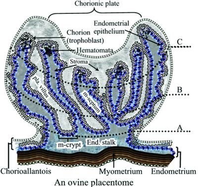
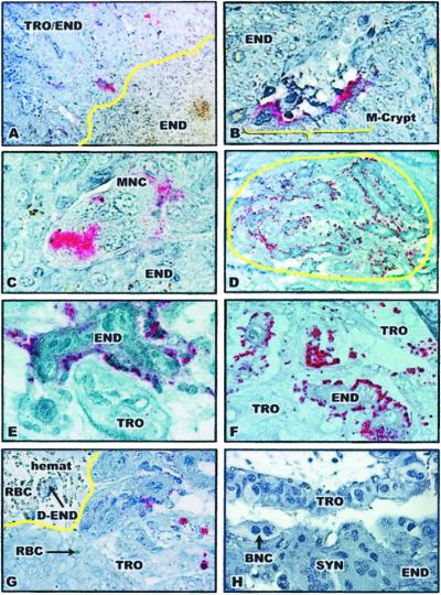
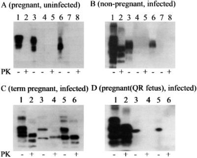
Ovine scrapie is a fatal neurodegenerative disorder that may be transmitted through exposure to infected uterine and placental tissues. Susceptibility to scrapie is primarily controlled by polymorphisms in the prion protein (PrP) gene. Scrapie in the U.S. Suffolk breed and in many breeds in Europe occurs in sheep homozygous for glutamine (171QQ), but rarely in sheep heterozygous for glutamine and arginine (171QR) or homozygous for arginine (171RR) at codon 171 of the PrP gene. This study demonstrated that accumulation of PrP(Sc) in uterine-placental epithelial cells in the placentome was determined by fetal PrP genotype and the pregnancy status of scrapie-infected ewes. PrP(Sc) was detected in 171QQ placentomes of infected ewes, but not in placentomes of infected ewes pregnant with 171QR conceptuses or in the non-pregnant uterus of infected ewes. The distribution of PrP(Sc) plaques in placentomes was temporally associated with stage of gestation. There was a tendency toward increased size and number of placentomal PrP(Sc) plaques from the endometrial stalk (maternal side) to chorionic plate (fetal side). These results indicate that accumulation of PrP(Sc) is eliminated or reduced to undetectable levels in reproductive and placental tissues if infected ewes are not pregnant or conceive conceptuses with a resistant PrP genotype.


Figures

Figure 1
Figure 1
A schematic illustration of a medial cross section of an ovine placentome. The placentome is a natural chimera, consisting of interdigitating uterine (placentomal or caruncular endometrium) and placental (placentomal or cotyledonary chorioallantois) tissues. The medial cross section of a placentome is divided into three zones: zones A, B, and C. Zone A contains maternal endometrial crypts and the distal ends of the cotyledonary villous tree and is located closest to the endometrial stalk and myometrium, zone B is the intermediate area, and zone C is the distal (relative to myometrium) portion of the placentome containing the distal ends of endometrial septae and the chorionic plate. Zone C is typically characterized by structures known as arcade system or hematomata. The placentomal endometrium consists of the uterine superficial epithelium and associated stroma, but no glands. Interplacentomal endometrium consists of loosely attached intercotyledonary chorioallantois and intercaruncular endometrium, which is rich in glands. Pla. villus, placental villus; m-septum, maternal endometrial septum; m-crypt, maternal crypt; end. stalk, endometrial stalk.
Figure 2
Figure 2
Immunohistochemical analysis of PrPSc in placentomes of scrapie-infected ewes. The presence of pink staining is indicative of the presence of PrPSc. TRO/END, trophoblast-endometrium interdigitation; TRO, trophoblast; END, endometrium; m-crypt, maternal crypt; MNC, multinucleated cells; RBC, maternal erythrocytes; hemat, hematomata; D-END, degenerating endometrium; ALL, allantoic cavity; BNC, binucleate cells; SYN, endometrial syncytium. (A) PrPSc plaques in the 171QQ placentome of a scrapie-infected ewe at day 40 of pregnancy (×100). PrPSc was seen only in endometrial/placental interdigitation, not in endometrium alone. The yellow line represents the border between maternal endometrium and trophoblast/endometrium interdigitation at the fetal–maternal interface at the bottom of the maternal crypt. (B) PrPSc in cells in the maternal crypt (m-crypt) in the 171QQ placentome of an early pregnant scrapie-infected ewe (×200). (C) PrPSc in a multinucleated cell (MNC) located in the maternal crypt in the 171QQ placentome of an early pregnant scrapie-infected ewe (×400). (D) A PrPSc plaque in zone C of a 171QQ placentome of a term pregnant scrapie-infected ewe (×100). The yellow circle defines the PrPSc plaque. (E) PrPSc in maternal endometrial epithelial cells in the 171QQ placentome of a term pregnant scrapie-infected ewe (×400). (F) PrPSc in both endometrial epithelial cells (END) and trophoblast cells (TRO) in the 171QQ placentome of a term pregnant scrapie-infected ewe (×200). (G) PrPSc in placental trophoblast cells (TRO) located along the hematomata (hemat) (×400). Phagocytosed RBCs (RBC and arrow) were apparent in the placental trophoblast cells. The yellow line defines part of the hematomata containing maternal blood (RBC) and degenerating endometrium (D-END). (H) A placentomal cross section near the endometrial stalk stained with an isotype-matched mAb (×400).
Figure 3
Figure 3
Western blot of PrPSc in tissue homogenates of brainstem, uterine caruncular endometrium, and placental cotyledonary chorioallantois of pregnant and non-pregnant ewes with or without scrapie. Protein from 0.5 mg brainstem or 30 mg endometrium, chorioallantois, and other tissues was loaded in each lane. PK (20 μg/ml) treatment at 37°C for 30 min of each tissue homogenate is indicated at the bottom of each panel (+). (A) Western blot of tissue homogenates prepared from brainstem (lanes 1 and 2), uterine caruncular endometrium (lanes 3–5), and cotyledonary chorioallantois (lanes 6–8) of an uninfected ewe (171QQ) pregnant (day 145 post mating) with a 171QQ conceptus. Samples in lanes 4, 5, 7, and 8 were prepared with sarkosyl and ultracentrifugation. Tissue homogenates were with (lanes 2, 5, and 8) or without (lanes 1, 3, 4, 6, and 7) PK treatment. (B) Western blot of tissue homogenates of brainstem (lanes 1 and 2), endometrium (lanes 3–5), and myometrium (lanes 6–8) of a non-pregnant scrapie-infected ewe (171QQ). Samples in lanes 4 and 5 and in lanes 7 and 8 were prepared with sarkosyl and ultracentrifugation. Tissue homogenates were with (lanes 2, 5, and 8) or without (lanes 1, 3, 4, 6, and 7) PK treatment. (C) Western blot of tissue homogenates prepared from brainstem, caruncular endometrium, and cotyledonary chorioallantois of a scrapie-infected ewe (177QQ) pregnant with a 171QQ conceptus. Lanes 1 and 2, brainstem prepared without sarkosyl and ultracentrifugation; lanes 3 and 4, uterine caruncular endometrium prepared with sarkosyl and ultracentrifugation; and lanes 5 and 6, placental cotyledonary chorioallantois prepared with sarkosyl and ultracentrifugation. Tissue homogenates were with (lanes 2, 4, and 6) or without (lanes 1, 3, and 5) PK treatment. (D) Western blot of tissue homogenates prepared from brainstem, caruncular endometrium, and cotyledonary chorioallantois of a scrapie-infected ewe (177QQ) pregnant with a 171QR conceptus. Lanes 1 and 2 (brainstem), prepared without sarkosyl and ultracentrifugation; lanes 3 and 4 (uterine caruncular endometrium), prepared with sarkosyl and ultracentrifugation; and lanes 5 and 6, placental cotyledonary chorioallantois prepared with sarkosyl and ultracentrifugation. Tissue homogenates were with (lanes 2, 4, and 6) or without (lanes 1, 3, and 5) PK treatment.
Figure 4
Figure 4
Distribution of PrPSc plaques in zones A, B, and C of the placentome of ewes infected with scrapie during early (n = 7) and term (n = 2) stages of pregnancy. The total number of PrPSc plaques in medial sections of two separate placentomes of each scrapie-infected ewe was counted microscopically. The results are expressed as the percentage of total PrPSc plaques.

References

    1. Prusiner S B. Science. 1982;216:136–144. - PubMed
    1. Oesch B, Westaway D, Walchli M, McKinley M P, Kent S B, Aebersold R, Barry R A, Tempst P, Teplow D B, Hood L E. Cell. 1985;40:735–746. - PubMed
    1. Basler K, Oesch B, Scott M, Westaway D, Walchli M, Groth D F, McKinley M P, Prusiner S B, Weissmann C. Cell. 1986;46:417–428. - PubMed
    1. Aguzzi A, Weissmann C. Haemophilia. 1998;4:619–627. - PubMed
    1. Prusiner S B. Proc Natl Acad Sci USA. 1998;95:13363–13383. - PMC - PubMed

Publication types