. 2002 Apr 30;99 Suppl 2(Suppl 2):6460-5.
doi: 10.1073/pnas.082526499. Epub 2002 Apr 16.

Segmented nanofibers of spider dragline silk: atomic force microscopy and single-molecule force spectroscopy

Affiliations

Segmented nanofibers of spider dragline silk: atomic force microscopy and single-molecule force spectroscopy

E Oroudjev et al. Proc Natl Acad Sci U S A. .

Abstract

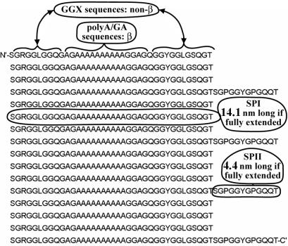
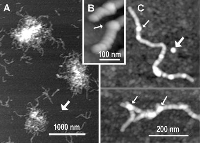
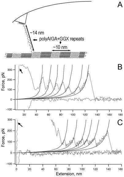
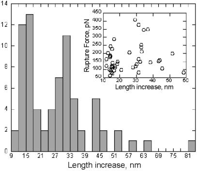
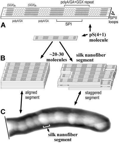
Despite its remarkable materials properties, the structure of spider dragline silk has remained unsolved. Results from two probe microscopy techniques provide new insights into the structure of spider dragline silk. A soluble synthetic protein from dragline silk spontaneously forms nanofibers, as observed by atomic force microscopy. These nanofibers have a segmented substructure. The segment length and amino acid sequence are consistent with a slab-like shape for individual silk protein molecules. The height and width of nanofiber segments suggest a stacking pattern of slab-like molecules in each nanofiber segment. This stacking pattern produces nano-crystals in an amorphous matrix, as observed previously by NMR and x-ray diffraction of spider dragline silk. The possible importance of nanofiber formation to native silk production is discussed. Force spectra for single molecules of the silk protein demonstrate that this protein unfolds through a number of rupture events, indicating a modular substructure within single silk protein molecules. A minimal unfolding module size is estimated to be around 14 nm, which corresponds to the extended length of a single repeated module, 38 amino acids long. The structure of this spider silk protein is distinctly different from the structures of other proteins that have been analyzed by single-molecule force spectroscopy, and the force spectra show correspondingly novel features.


Figures

Figure 1
Figure 1
The sequence of pS(4+1) recombinant silk protein, with selected SPI and SPII modules identified, as well as poly(A/GA) sequences and (GGX)n sequences. N′- and C'- indicate the N-terminal and C-terminal amino acids, respectively.
Figure 2
Figure 2
Silk nanofibers formed from pS(4+1) silk protein deposited on mica; AFM height images. (A) pS(4+1) silk protein is present primarily as aggregates of nanofibers. (B and C) Close-up AFM images of the pS(4+1) nanofibers show segmented substructure. Fat arrows indicate isolated blobs that are predicted to be segments of nanofibers, based on their sizes. Thin arrows indicate bulges, which often occur at branch points on nanofibers and may be due to nanofibers overlapping.
Figure 3
Figure 3
Single-molecule force spectroscopy for pS(4+1) silk protein molecules. (A) Diagram of experimental setup, in which rupture peaks are believed to stretch one or more poly(A/GA+GGX) repeating units. (B and C) Force spectra (force-vs.-extension curves) for the unfolding of single molecules of pS(4+1) silk protein. The WLC model curves are fitted to each rupture force peak. The persistence length for each fit is 0.4 nm. Arrows indicate high-force rupture events at the beginning of the pulls, most likely because of multiple protein molecules attached to the tip of the AFM probe.
Figure 4
Figure 4
Histogram analysis of length increase (peak-to-peak distances) for rupture events in pS(4+1) silk protein molecules. Note that most of the data coincide with ≈N × 14 nm in length increase, where N = 1, 2, or 3. (Inset) Length increase vs. corresponding rupture force.
Figure 5
Figure 5
A model for pS(4+1) silk nanofiber organization. (A) The single pS(4+1) protein molecule's polypeptide chain folds into a flat slab-like structure in which four hydrophobic β-sheets of poly(A/GA) (zig-zags) are separated by hydrophilic non-α-helical GGX structures (spirals). Four poly(A/GA) β-strands form each of the β-sheets that compose the crystalline-like structures of spider dragline silk. The (GGX)n sequences are random coils or 310-helices or other non-α-helical helical structures. (B) Approximately 30 of these slab-like molecules form a “stack” or “nanofiber segment” because of hydrophobic interactions between β-sheets in aqueous environment. These nanofiber segments are thought to bind to each other through specific “fiber-forming” signals/structures at their ends. Alternate segment models show either perfect or imperfect (staggered) alignment. (C) The whole pS(4+1) protein nanofiber can be viewed as a chain of segments with each segment representing a single pS(4+1) protein “stack.” Under a stretching force (not shown), as in the draw-down step of silk processing, the secondary structure of semiamorphous GGX “matrix” transitions into a more extended form and locks into a 31-helix or β-strand configuration. Numerous inter- and intramolecular H-bonds are formed at this point between these newly formed structures.

References

    1. Hinman M B, Jones J A, Lewis R V. Trends Biotechnol. 2000;18:374–379. - PubMed
    1. Hayashi C Y, Shipley N H, Lewis R V. Int J Biol Macromol. 1999;24:271–275. - PubMed
    1. Tirrell D A. Science. 1996;271:39–40. - PubMed
    1. Cunniff P M, Fossey S A, Auerbach M A, Song J W, Kaplan D L, Adams W W, Eby R K, Mahoney D, Vezie D L. Polym Adv Technol. 1994;5:401–410.
    1. Vollrath F, Knight D P. Nature (London) 2001;410:541–548. - PubMed

Publication types

LinkOut - more resources