. 2002 Apr 30;99(9):6358-63.
doi: 10.1073/pnas.082658499. Epub 2002 Apr 16.

Sustained stimulation shifts the mechanism of endocytosis from dynamin-1-dependent rapid endocytosis to clathrin- and dynamin-2-mediated slow endocytosis in chromaffin cells

Affiliations

Sustained stimulation shifts the mechanism of endocytosis from dynamin-1-dependent rapid endocytosis to clathrin- and dynamin-2-mediated slow endocytosis in chromaffin cells

Cristina R Artalejo et al. Proc Natl Acad Sci U S A. .

Erratum in

  • Proc Natl Acad Sci U S A 2002 Jun 25;99(13):9082

Abstract

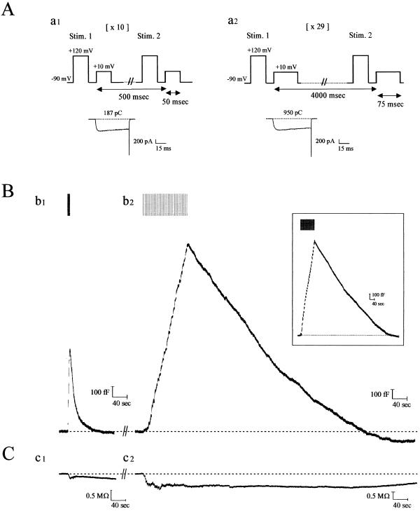
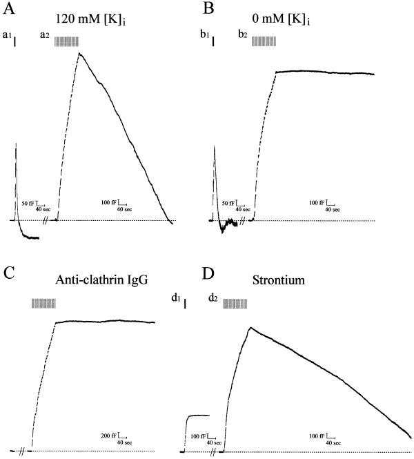
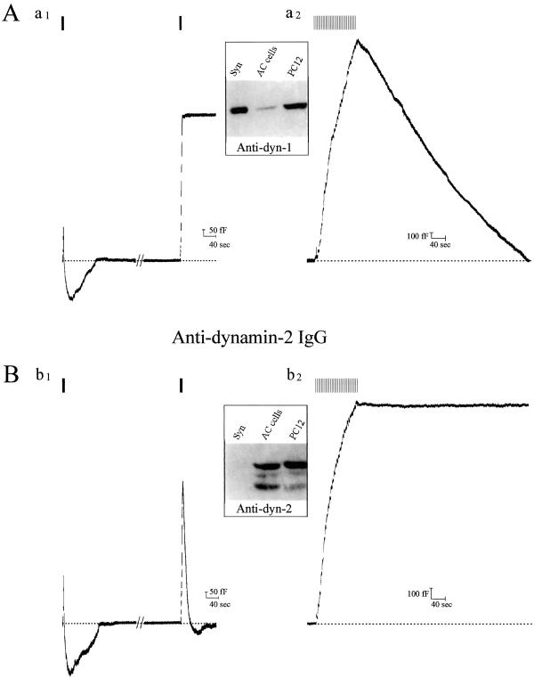
Transient stimulation of secretion in calf chromaffin cells is invariably followed by rapid endocytosis (RE), a clathrin- and K(+)-independent process with a half time of several seconds. Here we show that when exocytosis is triggered in a more sustained manner, a much slower form of endocytosis (SE) replaces RE. SE is complete within 10 min and is abolished when anticlathrin antibodies are introduced into the cell or when intracellular K(+) is removed. RE, but not SE, is blocked by intracellular administration of antidynamin-1 antibodies; the inverse specificity was found for antidynamin-2 antibodies. Replacement of extracellular Ca(2+) by Ba(2+) or Sr(2+) completely blocked RE but had little effect on SE. Thus chromaffin cells exhibit two kinetically and mechanistically distinct forms of endocytosis that are coupled to different extents of exocytosis and are mediated by different isoforms of dynamin. We surmise that RE is associated with the transient fusion ("kiss-and-run") mechanism of transmitter release and is the prevalent means of vesicle recapture and recycling under normal physiological conditions, whereas the clathrin-based SE mechanism comes into play only at higher levels of stimulation and may be associated with complete fusion of vesicles with the plasma membrane.


Figures

Figure 1
Figure 1
Kinetics of endocytosis are altered with degree of stimulation of calf chromaffin cells. (A) Patch-clamped calf chromaffin cells were stimulated with depolarizing pulse trains of 10 × 50 msec (with 0.5 sec between each stimulus; a1) or 29 × 75 msec (with 4 sec between each stimulus; a2). Before each depolarization, a prepulse was administered to recruit facilitation L-type Ca2+ channels in this preparation (9). Typical Ca2+ currents recorded from the first depolarizations in a1 and a2 are shown below the protocols (numbers indicate cumulative Ca2+ entry for each protocol; note this is 5.5-fold larger for the sustained versus the transient paradigm). (B) Continuous Cm recordings after formation of whole-cell configuration from a single calf chromaffin cell, sequentially stimulated with the two protocols illustrated in A. (b1) With transient stimulation, exocytosis (rising phase of Cm trace) was immediately followed by RE (falling phase of Cm trace). (b2) After a 12-min recovery period, during which baseline Cm was attained and remained stable, a second round of secretion was elicited with the sustained protocol. Under these conditions, endocytosis was slow, and Cm returned to baseline in ≈9 min (see Table 1 for average values). (Inset) SE was evoked during the first round of stimulation, 3 min after establishing whole-cell configuration. Results from a typical experiment are shown [mean values (n = 12) were peak current (676 ± 44 pA), current integral (767 ± 36 pC), total Cm increase (1,750 ± 125 fF), rate of endocytosis (134 ± 20 fF/min) and endocytosis duration (10 ± 0.6 min), respectively]. (C) Conductance traces accompanying the records in B show that this parameter does not change in parallel and thus does not significantly contaminate the Cm records. In total, >90% of the cells tested expressed both forms of endocytosis.
Figure 2
Figure 2
Slow endocytosis is mediated by a clathrin-coated vesicle pathway and is independent of Ca entry. Cm records from cells in which secretion was evoked as in Fig. 1 for both RE and SE. (A) Pipette solution contained 120 mM K+; SE ensues and is complete within ≈8 min. (B) Pipette solution contained 0 mM K+ (replaced by Cs-glutamate); SE is absent (b2), but RE is unaffected (b1). No significant difference in Cm rise between the sustained protocols in A and B was found (see Table 1 for average values of this and other parameters from these experiments). (C) Anticlathrin IgG (1 mg/ml) diffused into the cell from the patch pipette in the presence of 120 mM K+. Monoclonal anticlathrin IgG [×22] was dialyzed against internal pipette solution; 10 min was allowed to elapse between attaining a whole-cell patch and recording an exo/endocytotic cycle. Note that no SE takes place in the presence of the IgG (n = 18). RE (not shown) was unaffected by this maneuver, as previously described (9). Cm increase was similar to control. (D) Ca2+ dependence of RE but not SE. Bath Ca2+ was replaced by equimolar Sr2+, and stimulation was conducted to elicit either RE (d1) or SE (d2). RE was blocked, whereas SE continued and was complete within ≈15 min. No significant difference in Cm increase was found in Sr2+ versus Ca2+.
Figure 3
Figure 3
Dynamin-1 mediates RE, whereas dynamin-2 mediates SE. Cm records from cells in which either affinity-purified (A) antidynamin-1-specific IgG or (B) antidynamin-2-specific IgG, both at 1 mg/ml, were introduced into calf chromaffin cells followed by transient or sustained stimulation. In A, note that antidynamin-1 IgG inhibits RE (a1) but has no effect on SE (a2). In B, antidynamin-2 IgG has no effect on RE (b1) but blocks SE (b2). Note in Bb1 that two rounds of exocytosis/RE occur, whereas in Aa1 the second round of RE is blocked after the antibody has diffused into the cell (in the first round, RE is normal because insufficient antibody has diffused into the cell; the extent of the first exocytosis in Aa1 and Bb1 appears smaller because of simultaneous endocytosis that is largely absent in the second round). (Insets) Reactivity of antidynamin-1 (c1)- and -2 (c2)-specific antibodies with lysates from rat brain synaptosomes (Syn; 10 μg of protein); calf chromaffin (AC) cells (100 μg), and PC12 cells (100 μg); immunoblots were performed as described (9) and developed by using enhanced chemiluminescence.
Figure 4
Figure 4
Dynamin-1 and -2 are differentially distributed in calf chromaffin cells. Fixed chromaffin cells were double-labeled with antidynamin-1 (green)- and -2 (red)-specific IgGs with appropriate fluorescent secondary IgGs and viewed by confocal microscopy (Olympus FluoView). Series of single 0.5 μm sections through one cell (bottom of cell, Top) shown at Right with corresponding differential interference contrast microscopy images at Left (Bar = 2 μm). Note the minimal overlap (pixels registering for both signals) indicating the distinct distribution of the two proteins. Image representative of >1,000 sections from >50 different cells.

References

    1. Palfrey H C, Artalejo C R. Neuroscience. 1998;83:969–989. - PubMed
    1. Heuser J E, Reese T E. J Cell Biol. 1973;57:315–344. - PMC - PubMed
    1. Heuser J E. Cell Biol Int Rep. 1989;13:1063–1076. - PubMed
    1. De Camilli P, Slepnev V I, Shupliakov O, Brodin L. In: Synapses. Cowan W M, Sudhof T C, Stevens C F, editors. Baltimore, MD: Johns Hopkins Univ. Press; 2000. pp. 217–274.
    1. Ceccarelli B, Hurlbut W P, Mauro A. J Cell Biol. 1973;57:499–524. - PMC - PubMed

Publication types