. 2002 Apr 16;99(8):5139-43.
doi: 10.1073/pnas.072650799.

Fibronectin extension and unfolding within cell matrix fibrils controlled by cytoskeletal tension

Affiliations

Fibronectin extension and unfolding within cell matrix fibrils controlled by cytoskeletal tension

Gretchen Baneyx et al. Proc Natl Acad Sci U S A. .

Abstract

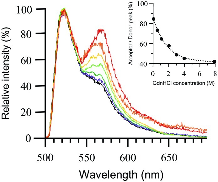
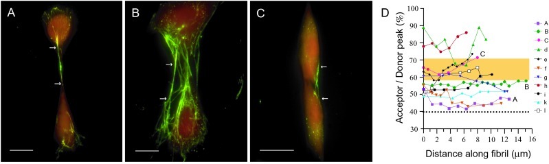
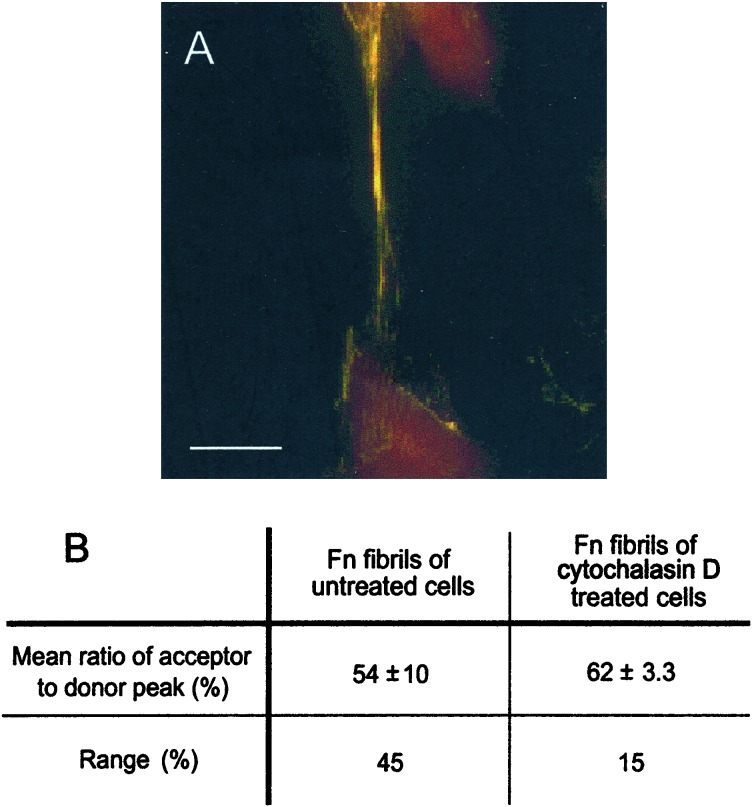
Evidence is emerging that mechanical stretching can alter the functional states of proteins. Fibronectin (Fn) is a large, extracellular matrix protein that is assembled by cells into elastic fibrils and subjected to contractile forces. Assembly into fibrils coincides with expression of biological recognition sites that are buried in Fn's soluble state. To investigate how supramolecular assembly of Fn into fibrillar matrix enables cells to mechanically regulate its structure, we used fluorescence resonance energy transfer (FRET) as an indicator of Fn conformation in the fibrillar matrix of NIH 3T3 fibroblasts. Fn was randomly labeled on amine residues with donor fluorophores and site-specifically labeled on cysteine residues in modules FnIII(7) and FnIII(15) with acceptor fluorophores. Intramolecular FRET was correlated with known structural changes of Fn in denaturing solution, then applied in cell culture as an indicator of Fn conformation within the matrix fibrils of NIH 3T3 fibroblasts. Based on the level of FRET, Fn in many fibrils was stretched by cells so that its dimer arms were extended and at least one FnIII module unfolded. When cytoskeletal tension was disrupted using cytochalasin D, FRET increased, indicating refolding of Fn within fibrils. These results suggest that cell-generated force is required to maintain Fn in partially unfolded conformations. The results support a model of Fn fibril elasticity based on unraveling and refolding of FnIII modules. We also observed variation of FRET between and along single fibrils, indicating variation in the degree of unfolding of Fn in fibrils. Molecular mechanisms by which mechanical force can alter the structure of Fn, converting tensile forces into biochemical cues, are discussed.


Figures

Figure 1
Figure 1
Sensitivity of energy transfer to Fn-D/A unfolding in denaturing solution. Fn-D/A emission spectra were measured by using an epifluorescence microscope with an attached spectrometer. Spectra were normalized to the donor peak so that changes in energy transfer were indicated by changes in the acceptor peak. Fn-D/A was progressively denatured over a range of Gdn⋅HCl concentrations, from 0 M to 8 M. Fn-D/A in 0 M Gdn⋅HCl is shown in red, 0.5 M in orange, 1 M in yellow, 2 M in light green, 3 M in dark green, 4 M in purple, and 8 M in black. The emission spectrum of Fn-D/A in 8 M Gdn⋅HCl was indistinguishable from that of a mixture of free donor and acceptor fluorophores in PBS. The small shoulder at 570 nm at 8 M Gdn⋅HCl was not caused by energy transfer, but by superposition of the donor fluorescence tail and fluorescence from direct excitation of the acceptor. (Inset) FRET (acceptor peak divided by donor peak) as a function of Gdn⋅HCl concentration.
Figure 2
Figure 2
Fluorescence images and spectra of Fn-D/A in fibroblast matrix fibrils. (AC) Fn-D/A was added to the culture medium of NIH 3T3 fibroblasts and incorporated into their fibrillar matrix. Excess unlabeled Fn was added to prevent intermolecular energy transfer. Fibrils visibly separated from other fibrils and from cells were analyzed. Arrows indicate beginning and end points of a series of spectra collected along a fibril. Spectra were integrated from 0.5-μm fibril segments spaced roughly 1 μm apart. (Scale bars, 10 μm.) (D) FRET varied between fibrils and along individual fibrils. Spectra labeled A, B, and C correspond to fibrils in images. Other fibrils that were analyzed are designated by lowercase letters, d–l. The shaded region indicates the range of FRET observed for Fn-D/A in fibrils of cells treated with cytochalasin D, a reagent that disrupts cytoskeletal tension (n = 78, 8 fibrils). The dashed line at the bottom indicates the level of FRET from Fn-D/A in 8 M Gdn⋅HCl in PBS.
Figure 3
Figure 3
Effect of cytochalasin D on conformation of Fn-D/A in matrix fibrils. (A) Fluorescence image of fibril after treatment of cells with 10 μM cytoD for 1 h after incorporation of Fn-D/A into fibrils. Fibrils in cell samples treated with cytoD appeared predominantly yellow, indicating a higher level of FRET than in the predominantly green fibrils of untreated samples. The increase in FRET indicated that cytoD allowed refolding of Fn-D/A in fibrils relative to untreated samples. (Scale bar, 10 μm.) (B) The mean level of FRET (acceptor peak divided by donor peak) from fibrils in untreated samples, 54% ± 10% (n = 115, 11 fibrils), was significantly lower than in samples treated with cytoD, 62 ± 3.3% (n = 78,8 fibrils) (P < 0.001, two-sample t test). The range of FRET in fibrils of cytoD-treated cells (15%) was also narrower than that of untreated cells (45%).

References

    1. Sechler J L, Schwarzbauer J E. J Biol Chem. 1998;273:25533–25536. - PubMed
    1. Sechler J L, Corbett S A, Wenk M B, Schwarzbauer J E. Ann NY Acad Sci. 1998;857:143–154. - PubMed
    1. Darribere T, Schwarzbauer J E. Mech Dev. 2000;92:239–250. - PubMed
    1. Leahy D J, Aukhil I, Erickson H P. Cell. 1996;84:155–164. - PubMed
    1. Spitzfaden C, Grant R P, Mardon H J, Campbell I D. J Mol Biol. 1997;265:565–579. - PubMed

Publication types

LinkOut - more resources