. 2002 Apr 16;99(8):5367-72.
doi: 10.1073/pnas.082117899.

Structural basis for recruitment of CBP/p300 by hypoxia-inducible factor-1 alpha

Affiliations

Structural basis for recruitment of CBP/p300 by hypoxia-inducible factor-1 alpha

Steven J Freedman et al. Proc Natl Acad Sci U S A. .

Abstract

Adaptation to hypoxia is mediated by transactivation of hypoxia-responsive genes by hypoxia-inducible factor-1 (HIF-1) in complex with the CBP and p300 transcriptional coactivators. We report the solution structure of the cysteine/histidine-rich 1 (CH1) domain of p300 bound to the C-terminal transactivation domain of HIF-1 alpha. CH1 has a triangular geometry composed of four alpha-helices with three intervening Zn(2+)-coordinating centers. CH1 serves as a scaffold for folding of the HIF-1 alpha C-terminal transactivation domain, which forms a vise-like clamp on the CH1 domain that is stabilized by extensive hydrophobic and polar interactions. The structure reveals the mechanism of specific recognition of p300 by HIF-1 alpha, and shows how HIF-1 alpha transactivation is regulated by asparagine hydroxylation.


Figures

Figure 1
Figure 1
Domain structures and sequence alignments of HIF-1α and p300 and structure of the CTAD/CH1 complex. (a) Functional domains of CBP/p300 (Upper) and HIF-1α (Lower). Domains in CBP/p300 are nuclear hormone receptor-binding domain (Nu), cysteine/histidine-rich domains (CH1, CH2, and CH3), CREB-binding domain (KIX), bromodomain (Br), histone acetyltransferase domain (HAT), glutamine-rich domain (Q), and IRF-3-binding domain (I). The CH1 and CH3 domains are structurally homologous and also have been termed TAZ1 and TAZ2, respectively. Domains in HIF-1α are basic helix-loop-helix domain (bHLH), Per-Arnt-Sim homology domain (PAS), and N- and C-terminal transactivation domains (NTAD, CTAD). Transcription factors that have been shown to associate with CBP/p300 are shown above the interacting domains. Those that have been structurally characterized in complex with their respective CBP/p300 binding domains are highlighted in red. The domains of p300 and HIF-1α that form the complex studied here are highlighted in dark blue and red, respectively. This figure was adapted from Vo and Goodman, 2001 (16). (b) The sequence of the human HIF-1α CTAD used for structure determination (top line) is aligned with the homologous regions of HIF-1α from other species and human HIF-2α. Note that the structured portion of HIF-1α (residues 792–824) is nearly 100% conserved. (b and c) Elements of secondary structure are indicated above the alignment. The shaded vertical bars above the alignment indicate the fraction of the residue surface that is buried in the HIF-1α/p300 complex interface. (c) The sequence of human p300 CH1 used for structure determination (top line) is aligned with the homologous regions of p300 and CBP. The single histidine and three cysteines that form each of the three strictly conserved Zn2+-binding sites are shaded violet, green, or yellow. Residues highlighted in blue are conserved residues that form the hydrophobic core of the human CH1 structure. Most of these residues are conserved in the CH3 domain. The residues that are buried in the interface between the HIF-1α CTAD and p300 CH1 domain are distributed among all four helices but are most prominent along α3. The arrowheads under the alignment indicate the positions of insertions relative to the human p300 CH1 sequence. The number of residues inserted in the CH3 domains are indicated; those numbers with asterisks are insertions in the C. elegans sequence of CBP CH1. The aligned sequences are: h, Homo sapiens; b, Bos taurus; m, Mus musculus; x, Xenopus laevis; d, Drosophila melanogaster; c, Caenorhabditis elegans. (d) Stereoview of 17 superimposed CTAD/CH1 complex structures. (e) Ribbon diagram of the lowest-energy CTAD/CH1 structure. The fold of CH1 (royal blue/light blue) and CTAD (red/orange) is described in the text. Helices α1 (residues 332–354), α2 (residues 367–379), α3 (residues 391–405), and α4 (residues 414–418) refer to the α-helical regions of p300 CH1; residues 332–334 are 3:10 helix. Helices αA (residues 797–803) and αB (residues 816–822) refer to the α-helical regions of the HIF-1α CTAD; residues 815–817 are 3:10 helix. Green spheres indicate the three putative Zn2+ ions in CH1 and are labeled Zn1 through Zn3. (f) Superposition of the CH1 domain from the CTAD/CH1 complex with the free CBP CH3 domain (25). Note the similar folds from α1 through α3 (gray) and the conformational differences of the third Zn2+-binding turn and α4 (CH1 residues 407–418 in blue and CH3 residues 1835–1850 in yellow). The eight-residue insertion (residues 353–363) in the first Zn2+-binding turn of CH1 relative to the homologous region of CH3 (residues 1788–1790) is similarly color-coded. d was prepared with MOLMOL (40), e was prepared with MOLSCRIPT (41), and f was prepared with INSIGHTII (Accelrys).
Figure 2
Figure 2
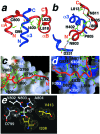
Intermolecular contacts between the HIF-1α CTAD and p300 CH1 domains. (a) A region of the CTAD/CH1 complex is magnified to illustrate some of the important hydrophobic contacts that define the topology of the interaction. CTAD wraps around CH1 like a clamp such that αA and αB rest in grooves on either side of α3. Note the parallel configuration of the CH1 helix sandwiched between the two CTAD helices. Several hydrophobic side chains considered to contribute to the binding energy are displayed as sticks and are labeled by residue and number. (b) A similar region of the complex (interhelical loop of CTAD, N terminus of α1 and α3 from CH1) is shown in a different orientation to illustrate putative intermolecular hydrogen bond contacts that stabilize the complex. The presence of hydrogen bonds is supported by NOE and structure analyses. (c and d) The N- and C-terminal regions of the HIF-1α CTAD (red ribbon) are shown with the p300 CH1 domain represented as an accessible surface. The surface is colored by charge and is scaled from −10 kT/e (red) to +10 kT/e (blue). Selected HIF-1α side chains are labeled in black. Basic residues in CH1 are labeled in white. (e) Position of Asn-803 in the CTAD/CH1 complex indicates how β-hydroxylation would inhibit binding. CH1 residues are shown in yellow and CTAD residues are shown in white. The Asn-803 Hβ (pro-R) and Hβ (pro-S) are colored green. Van der Waals surfaces are shown for residues surrounding the Asn-803 side chain. Substitution of either the pro-R or pro-S β-protons with a hydroxyl group would disfavor complex formation because of steric and hydrogen-bonding considerations (see text). (a, b, and e) Complexes were prepared with INSIGHTII (Accelrys). (c and d) Complexes were prepared with GRASP (42).
Figure 3
Figure 3
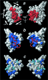
The HIF-1α CTAD/p300 CH1 interface. (ac) The CH1 domain is shown in the same orientation, and the view on the right is rotated ≈180° about the vertical axis. (a) A space-filling CPK model of the complex. The HIF-1α CTAD is colored red, and the p300 CH1 domain is shown in white. The HIF-1α CTAD is embedded in the CH1 scaffold so that the complex appears as a single domain. (b) The surface of the CH1 domain is shown in white with the HIF-1α CTAD-binding surface shaded blue. The bound HIF-1α CTAD is shown as a red ribbon. (c) A space-filling model of CH1 in which residues that are identically conserved in human CBP/p300 CH3 are shown in royal blue, and those that are conservatively substituted are shown in light blue. Comparison of b and c shows that the HIF-1α-binding surface is not well conserved in the CH3 domain, which binds different transactivation domains. This figure was prepared with MOLMOL (40).

References

    1. Semenza G L. Annu Rev Cell Dev Biol. 1999;15:551–578. - PubMed
    1. Iyer N V, Kotch L E, Agani F, Leung S W, Laughner E, Wenger R H, Gassmann M, Gearhart J D, Lawler A M, Yu A Y, et al. Genes Dev. 1998;12:149–162. - PMC - PubMed
    1. Semenza G L. Trends Mol Med. 2001;7:345–350. - PubMed
    1. Salceda S, Caro J. J Biol Chem. 1997;272:22642–22647. - PubMed
    1. Ivan M, Kondo K, Yang H, Kim W, Valiando J, Ohh M, Salic A, Asara J M, Lane W S, Kaelin W G., Jr Science. 2001;292:464–468. - PubMed

Publication types

MeSH terms

Associated data