. 2002 Apr 16;99(8):5567-72.
doi: 10.1073/pnas.082100399.

IRAK-4: a novel member of the IRAK family with the properties of an IRAK-kinase

Affiliations

IRAK-4: a novel member of the IRAK family with the properties of an IRAK-kinase

Shyun Li et al. Proc Natl Acad Sci U S A. .

Abstract

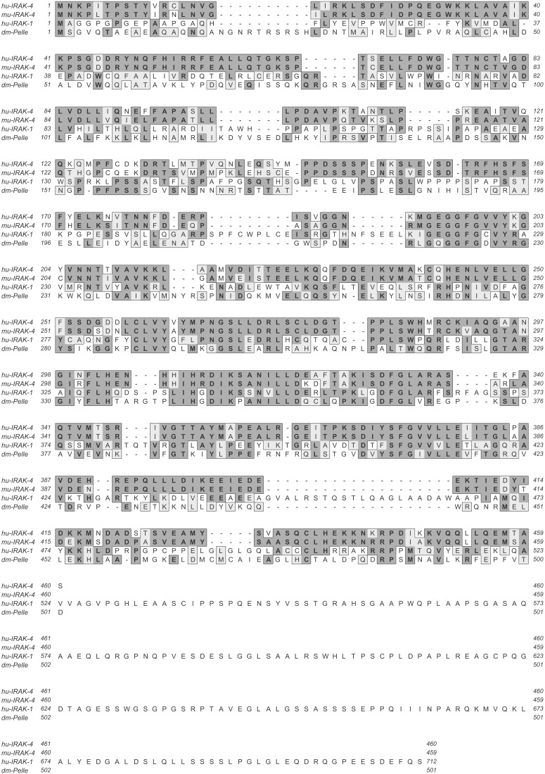
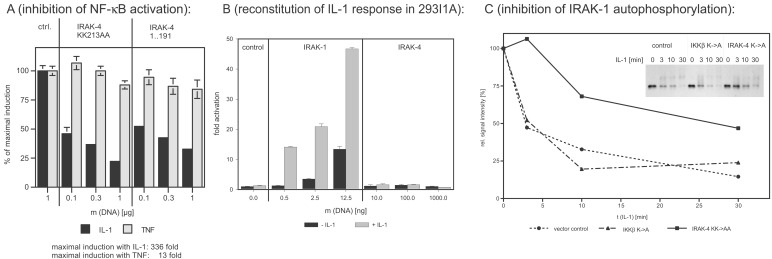
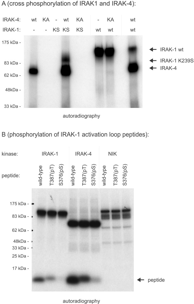
Toll/IL-1 receptor family members are central components of host defense mechanisms in a variety of species. One well conserved element in their signal transduction is Ser/Thr kinases, which couple early signaling events in a receptor complex at the plasma membrane to larger signalosomes in the cytosol. The fruit fly Drosophila melanogaster has one member of this family of kinases, termed Pelle. The complexity of this pathway is vastly increased in vertebrates, and several Pelle homologs have been described and termed IL-1 receptor-associated kinase (IRAK). Here we report the identification of a novel and distinct member of the IRAK family, IRAK-4. IRAK-4 is the closest human homolog to Pelle. Endogenous IRAK-4 interacts with IRAK-1 and TRAF6 in an IL-1-dependent manner, and overexpression of IRAK-4 can activate NF-kappa B as well as mitogen-activated protein (MAP) kinase pathways. Most strikingly, and in contrast to the other IRAKs, IRAK-4 depends on its kinase activity to activate NF-kappa B. In addition, IRAK-4 is able to phosphorylate IRAK-1, and overexpression of dominant-negative IRAK-4 is blocking the IL-1-induced activation and modification of IRAK-1, suggesting a role of IRAK-4 as a central element in the early signal transduction of Toll/IL-1 receptors, upstream of IRAK-1.


Figures

Figure 1
Figure 1
Protein sequences of human IRAK-4, murine IRAK-4, human IRAK-1, and Drosophila melanogaster Pelle. Dark and light shading indicates identical and similar residues, respectively.
Figure 2
Figure 2
Interaction of IRAK-4 with components of the IL-1R complex. (A) Coprecipitation of MyD88 with IRAK-4 and IRAK-1. The 293 cells were transfected with expression plasmids coding for myc-tagged IRAK-1 or IRAK-4 (each wild-type and kinase-inactive mutant) alone or together with Flag-tagged MyD88. MyD88 was immunoprecipitated (IP) with anti-Flag mAb, and coprecipitating IRAKs were detected with anti-myc antiserum (Top). Western blot analysis (WB) of the same blot with mAb to the Flag epitope shows that similar amounts of MyD88 were IP (Middle). The lysates of the transfected cells were immunoblotted with myc antiserum to monitor the expression of IRAKs (Bottom). (B) Coprecipitation of TRAF6 with IRAK-4 and IRAK-1. The 293 cells were transfected with expression plasmids coding for myc-tagged IRAK-1 or IRAK-4 alone or together with Flag-tagged TRAF6. IP and WB were performed as described in A. (C) IL-1-induced coprecipitation of IRAK-4 with TRAF6 and IRAK-1. Untransfected 293RI cells (2 × 10⁸ cells per sample) were stimulated for the indicated amount of time with 100 ng/ml of IL-1β. Cell lysates were IP with either IRAK-4 antiserum or preimmune serum. Coprecipitating IRAK-1 and TRAF6 were detected with polyclonal antisera (Upper). In a second Western blot, the same membrane was used to show equal precipitation of IRAK-4.
Figure 3
Figure 3
Activation of Elk1, cJun, and NF-κB by IRAK-4. (A) Elk1 activation. The 293 cells were transfected with the indicated amounts of expression plasmids for IRAK-4 and MEK1 (positive control), pRSVβgal for normalization, the GAL4-dependent reporter construct pFR-luc and the transactivator plasmid pFA2-Elk1, a fusion of the DNA binding domain of GAL4 and the transactivation domain of Elk1. The y axis represents the normalized fold of luciferase activity induction relative to cells transfected with a transactivator plasmid lacking the transactivation domain. (B) cJun activation. The experiment was performed as described above, with expression plasmids for MEKK as positive control and pFA2-cJun as transactivator. (C) Activation of NF-κB. The 293 cells were transfected with an NF-κB-dependent ELAM-luc reporter gene construct, pRSVβgal for normalization, and the indicated amounts of wild-type IRAK-4, kinase-inactive IRAK-4 (IRAK-4 KK213AA), and truncated IRAK-4 (amino acids 1–191). Twenty-four hours after transfection, luciferase and β-galactosidase activity were determined. The y axis represents the normalized fold of luciferase activity induction relative to cells transfected with empty vector.
Figure 4
Figure 4
Dominant-negative effects of IRAK-4 mutants and reconstitution experiments in IRAK-1-deficient 293 cells. (A) Inhibition of IL-1-induced NF-κB activation. The 293RI cells were transfected with an NF-κB-dependent ELAM-luc reporter gene construct, pRSVβgal for normalization, and the indicated amounts of kinase-inactive IRAK-4 (IRAK-4 KK213AA) and truncated IRAK-4 (amino acids 1–191). Twenty-four hours after transfection, cells were stimulated with 50 ng/ml of IL-1β or 100 ng/ml of tumor necrosis factor α for 6 h, and luciferase and β-galactosidase activity were determined. The y axis represents the percentage of NF-κB activation relative to cells transfected with empty vector. (B) Reconstitution of IL-1 response in 293I1A. IRAK-1-deficient 293 cells [293I1A(16)] were transfected and stimulated as described for Fig. 4A. The y axis represents the fold of NF-κB activation relative to cells transfected with empty vector. (C) Inhibition of IL-1-induced IRAK-1 activation. The 293RI cells were transfected with control vector, kinase-inactive IKKβ (negative control), or kinase-inactive IRAK-4 for 24 h. After stimulation with 100 ng/ml of IL-1β for the indicated amount of time, the cells were lysed, and IRAK-1 was immunoprecipitated with the mAb 2A9. The activation level of IRAK-1 was assessed by determining the shift in apparent molecular weight and subsequent degradation of IRAK-1 by Western blotting (Inset). The signal intensity was quantified by densitometry and plotted as a function of incubation time.
Figure 5
Figure 5
Phosphorylation of the IRAK-1 activation loop. (A) Crossphosphorylation of IRAK-1 and IRAK-4. Full-length IRAK-1 and IRAK-4 expressed in insect cells were tested for their ability to phosphorylate each other in vitro. One hundred nanograms of wild-type protein (wt) were incubated with kinase-inactive mutants (KA, IRAK-4 KK213AA; KS, IRAK-1 K239S) in the presence of [γ-32P]ATP. (B) Phosphorylation of IRAK-1 activation loop peptides. Synthetic peptides were derived from the IRAK-1 activation loop (amino acids 359–389). The substrate quality of wild-type peptides was compared with peptides, in which either Thr-387 or Ser-376 was replaced with phospho-threonine or phospho-serine, respectively. After incubation in an in vitro kinase assay with recombinant IRAK-1, recombinant IRAK-4, and recombinant NIK (negative control), phosphorylation of peptides was determined by incorporation of radioactive phosphate after SDS/PAGE and autoradiography.

References

    1. Kollewe, C. & Martin, M. U. (2002) Signal Trans., in press.
    1. Wesche, H. & Martin, M. U. (2002) Biochim. Biophys. Acta, in press. - PubMed
    1. Dinarello C A. Blood. 1996;87:2095–2147. - PubMed
    1. Fallon P G, Allen R L, Rich T. Trends Immunol. 2001;22:63–66. - PubMed
    1. Ulevitch R J, Tobias P S. Curr Opin Immunol. 1999;11:19–22. - PubMed

MeSH terms

Associated data