. 2002 May;50(5):617-28.
doi: 10.1177/002215540205000503.

Localization of caveolin 1 in aortic valve endothelial cells using antigen retrieval

Affiliations

Localization of caveolin 1 in aortic valve endothelial cells using antigen retrieval

Nalini M Rajamannan et al. J Histochem Cytochem. 2002 May.

Abstract

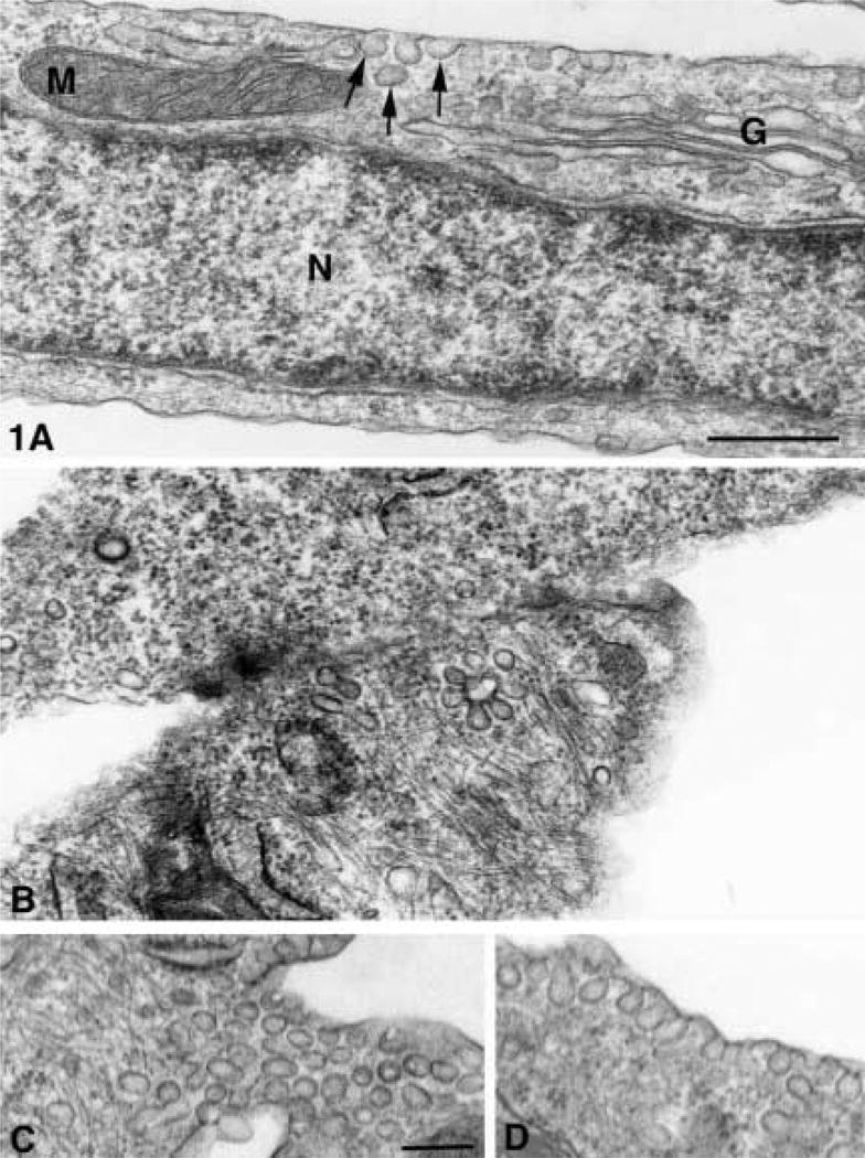
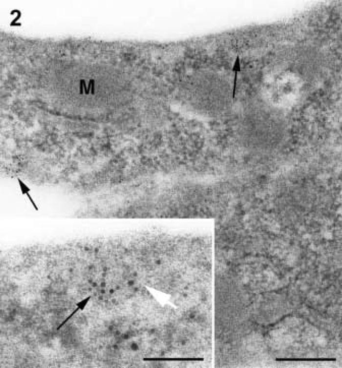
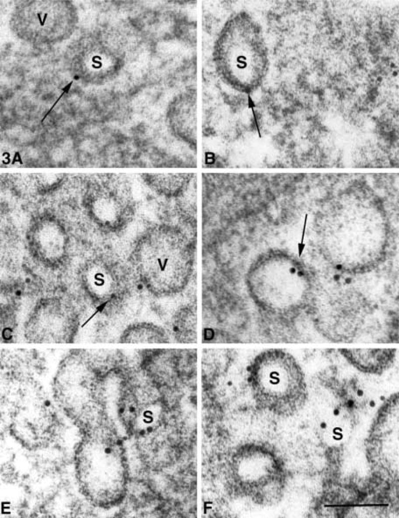
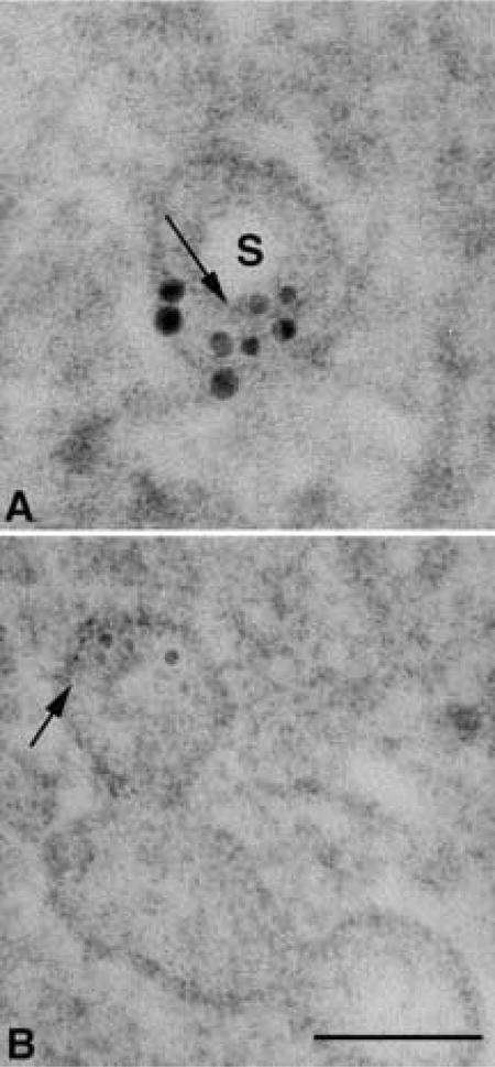
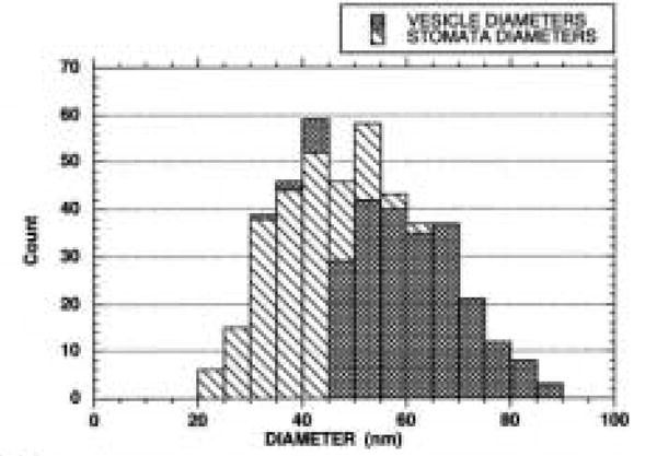
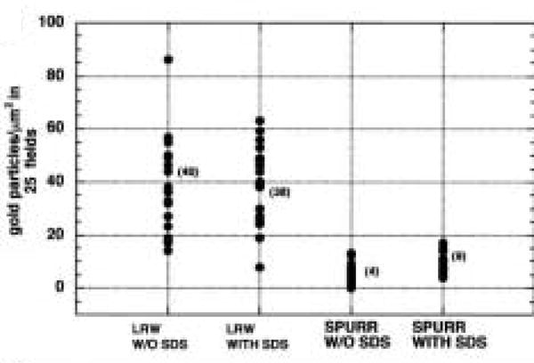
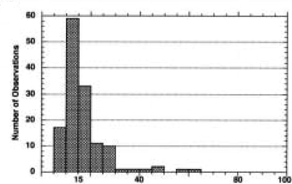
Ultrastructural analysis of aortic valve endothelial cells subjected to growth arrest revealed many vesicles defined as caveolae by the localization of caveolin. Translocation of caveolin after exposure to oxidized LDL suggests that the localization of caveolin may be a valuable tool to study models of early atherogenesis. In this study, several antigen retrieval protocols were tested in osmium-fixed and Spurr-embedded cells to determine the optimal method of antigen retrieval in our model system. SDS produced the most consistent labeling pattern. A quantitative evaluation revealed that SDS significantly increased the labeling density in Spurr-embedded cells. The labeling pattern appeared as clusters of gold particles, 15-40 nm in diameter, that were associated with membranes of a similar size which may represent the neck region of the caveolae.


Figures

Figure 1
Figure 1
(A) Normal cultured AVECs from pig with ultrastructural detail that can be demonstrated in sections osmium-fixed, uranyl en bloc-stained, and embedded in Spurr resin. Membranes of mitochondria (M), Golgi (G), caveolae (arrows), and those surrounding nucleus (N) show trilaminar structure of lipid bilayers. Bar = 400 nm. (BD) Serum-free aortic valve endothelial cell with VVO (arrow) in the border between two endothelial cells (B) and caveolae groups (arrow) aligned parallel to (C) and caveolae oriented perpendicular to the plasma membrane (D). Bar = 100 nm.
Figure 2
Figure 2
Serum-free AVEC without osmium and embedded in LR White resin. Membranes of mitochondria (M), caveolae, and plasmalemma have little contrast and are difficult to resolve. The labeling of caveolin (arrows) is patchy and appears to be concentrated in zones near the plasma membranes. Bar = 100 nm. (Inset) Higher magnification of gold labeling (black arrow) in LR White-embedded cells that have no postfixation in osmium showing membranes of reverse contrast (white arrow). Bar = 33 nm.
Figure 3
Figure 3
(AF) Caveolin labeling patterns in AVECs under serum-free conditions, embedded in Spurr resin and subjected to pre-labeling AR in SDS. The gold label (arrows) is consistently associated with vesicle membranes that surround the dense vesicle matrix (V) and clear areas (S) that often do not show a central dense knob. These smaller vesicle membranes may represent the neck region of the vesicle when the diaphragm has been removed in sectioning. Bar = 60 nm.
Figure 4
Figure 4
High magnification of two vesicles that appear to be sectioned through the stoma area. (A) In this section, the dense knob is not apparent and the stoma (S) is a clear area. Two semicircular gold clusters may represent the two angles of membrane curvature that mark the entrance into the vesicle through the neck region. (B) Labeling over small radiating tubular structures (arrow) with a diameter smaller than the 5-nm gold particles that may be associated with a dense central knob of the diaphragm. Arrow in A points to a cross-section of a tubular structure that corresponds with the diameter of the longitudinally oriented tubules demonstrated in B. Bar = 75 nm.
Figure 5
Figure 5
Distribution of vesicle diameters that define either dense vesicle matrix or clear area within caveolae. Most stomata diameters range from 20 to 40 nm in diameter, while those of the denser vesicle matrix range from 40 to 70 nm in diameter.
Figure 6
Figure 6
In LR White- and Spurr-embedded AVECs under serum-free conditions, a comparison of gold-labeling with and without antigen retrieval in SDS. LR White labeling is increased compared to Spurr labeling but there are no differences with and without SDS antigen retrieval. Labeling intensity after Spurr embedding is considerably less than labeling in LR White, but the signal increases after SDS antigen retrieval. The (mean) labeling density in each group is included.
Figure 7
Figure 7
Distribution of interpoint distances measured between a gold particle and the nearest neighboring gold particle within a gold cluster. Aggregation of 5-nm gold particles due to antibody and gold interactions may occur at distances up to 15 nm, but clusters of particles between 15 and 40 nm may suggest that the clusters are localizing in the neck areas of the vesicles where the membranes are smaller in diameter.

References

    1. Amino K, Honda Y, Ide C, Fujimoto T. Distribution of plasmalemmal Ca(2+)-pump and caveolin in the corneal epithelium during the wound healing process. Curr Eye Res. 1997;16:1088–1095. - PubMed
    1. Bendayan M, Zollinger M. Ultrastructural localization of antigenic sites on osmium-fixed tissues applying the protein A-gold technique. J Histochem Cytochem. 1983;31:101–109. - PubMed
    1. Blair A, Shaul PW, Yahanna IS, Conrad PA, Smart EJ. Oxidized low density lipoprotein displaces endothelial nitric-oxide synthase (eNOS) from plasmalemmal caveolae and impairs eNOS activation. J Biol Chem. 1999;274:32512–32519. - PubMed
    1. Brorson SH. Fixative-dependent increase in immunogold labeling following antigen retrieval on acrylic and epoxy sections. Biotech Histochem. 1999;74:248–260. - PubMed
    1. Brown D, Lydon J, McLaughlin M, Stuart–Tilley A, Tyszkowski R, Alper S. Antigen retrieval in cryostat sections and cultured cells by treatment with sodium dodecyl sulfate (SDS) Histochem Cell Biol. 1996;105:261–267. - PubMed