Comparative Study
. 2002 May;76(10):4773-84.
doi: 10.1128/jvi.76.10.4773-4784.2002.

Mutations in the yellow fever virus nonstructural protein NS2A selectively block production of infectious particles

Affiliations
Comparative Study

Mutations in the yellow fever virus nonstructural protein NS2A selectively block production of infectious particles

Beate M Kümmerer et al. J Virol. 2002 May.

Abstract

Little is known about the function of flavivirus nonstructural protein NS2A. Two forms of NS2A are found in yellow fever virus-infected cells. Full-length NS2A (224 amino acids) is the product of cleavage at the NS1/2A and NS2A/2B sites. NS2Aalpha, a C-terminally truncated form of 190 amino acids, results from partial cleavage by the viral NS2B-3 serine protease at the sequence QK /T within NS2A. Exchange of serine for lysine at this site (QKT-->QST) blocks the production of both NS2Aalpha and infectious virus. The present study reveals that this defect is not at the level of RNA replication. Despite normal structural region processing, infectious particles containing genome RNA and capsid protein were not released from cells transfected with the mutant RNA. Nevertheless, production of subviral prM/M- and E-containing particles was unimpaired. The NS2A defect could be complemented in trans by providing NS1-2A or NS1-2Aalpha. However, trans complementation was not observed when the C-terminal lysine of NS1-2Aalpha was replaced with serine. In addition to true reversions, NS2Aalpha cleavage site mutations could be suppressed by two classes of second-site changes. The first class consisted of insertions at the NS2Aalpha cleavage site that restored its basic character and cleavability. A second class of suppressors occurred in the NS3 helicase domain, in which NS3 aspartate 343 was replaced with an uncharged residue (either valine, alanine, or glycine). These mutations in NS3 restored infectious-virus production in the absence of cleavage at the mutant NS2Aalpha site. Taken together, our results reveal an unexpected role for NS2A and NS3 in the assembly and/or release of infectious flavivirus particles.


Figures

FIG. 1.
FIG. 1.
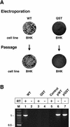
Phenotype of the YF NS2Aα QST mutant. (A) NS2A processing and the NS2Aα cleavage site. Shown is a diagram of the YF polyprotein encompassing NS2A. In addition to full-length NS2A, a C-terminally truncated form (NS2Aα) is produced. Cleavages to generate the C termini of NS2A and NS2Aα are mediated by the NS2B-NS3 serine protease (↓). The NS2Aα cleavage site is defined by the sequence QK↓T. (B) RNA analysis. BHK cells were electroporated with WT or NS2Aα QST mutant RNA transcripts. From 20 to 28 h postelectroporation, the viral RNA was labeled with [3H]uridine in the presence of actinomycin D. Total RNA was isolated, denatured, and resolved by electrophoresis through a 1% agarose gel. The gel was treated for fluorography, dried, and exposed to X-ray film. (C) Infectious-center assay. BHK cells were electroporated with the WT or mutant (QST) transcript, and serial 10-fold dilutions were seeded on 35-mm-diameter dishes in the presence of untransfected cells and overlaid with agarose. After 3 days, cells were fixed and crystal violet staining was performed. The values below the dishes indicate the amounts (PFU per microgram) of in vitro-transcribed RNA. (D) NS2A processing. BHK cells were metabolically labeled 20 to 25 h after electroporation with [35S]methionine-cysteine. YF-specific proteins were immunoprecipitated from cellular extracts with YF-specific HIAF. Proteins were solubilized, denatured, and resolved by electrophoresis through an SDS-12% polyacrylamide gel (12). The values on the left are molecular masses of standard proteins in kilodaltons.
FIG. 2.
FIG. 2.
Failure of the NS2Aα QST mutant to produce infectious virus. (A) Infectious-center and infectivity assays. WT or NS2Aα QST RNA transcripts were electroporated into BHK cells. Serial 10-fold dilutions were seeded on 35-mm-diameter dishes in the presence of the respective untransfected cells and overlaid with agarose. After 3 days, cells were fixed and crystal violet staining was performed. Supernatants from parallel liquid cultures were used to perform a plaque assay on new BHK cells. In addition, total cellular RNA was extracted from parallel cultures of reinfected cells and used for RT-PCR analysis. (B) RNA analysis. The region containing the NS2Aα cleavage site sequence was amplified from total cellular RNAs from the experiment described in panel A. As a control for DNA carryover, parallel reactions without reverse transcriptase (RT) were performed (lanes 2 and 4). Amplifications from the WT full-length plasmid (pWT) or the mutant full-length plasmid (pQST) served as positive controls (lanes 6 and 7). Lane M, DNA size markers (molecular sizes are in kilobases on the left). Control, no-template control.
FIG. 3.
FIG. 3.
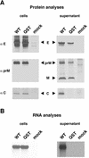
Processing and release of structural proteins and viral RNA from WT- and NS2Aα QST-transfected cells. (A) Structural protein analysis. BHK cells were metabolically labeled 20 to 25 h postelectroporation with [35S]methionine-cysteine. YF-specific proteins were precipitated from either cellular extracts or supernatants with polyclonal rabbit sera against E, prM, or C. Precipitated proteins were resolved on a Tricine-SDS-10% (αE) or -14% (αprM and αC) polyacrylamide gel (28). (B) RNA analysis. BHK cells were labeled 15 to 22 h postelectroporation with [3H]uridine in the presence of actinomycin D. RNA was extracted either from the cells or from virus pelleted from the supernatant. The extracted RNA was resolved on an agarose gel under denaturing conditions.
FIG. 4.
FIG. 4.
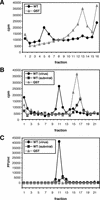
Characterization of particles released from WT- and NS2Aα QST-transfected cells. BHK cells were transfected with WT or NS2Aα QST mutant (QST) RNA transcripts, metabolically labeled with [35S]methionine-cysteine from 14 to 26 h posttransfection, concentrated by pelleting, and resuspended in buffer as described in Materials and Methods. (A) Rate-zonal sedimentation. WT or NS2Aα QST mutant particles were sedimented in parallel glycerol gradients as described in Materials and Methods. (B) Equilibrium banding. A portion of pooled fractions 5 and 6 [WT (virus)] and a portion of pooled fractions 12 to 14 [WT (subviral)] from the glycerol gradient shown in panel A were loaded on parallel tartrate gradients. For the NS2Aα QST mutant (QST), fractions 12 to 14 were pooled and analyzed. (C) Infectivity of the gradient fractions. Fractions of the gradients shown in panel B were analyzed for infectivity by plaque assay on BHK cells. In all panels, fractions are numbered from the bottom to the top.
FIG. 5.
FIG. 5.
trans-acting factors required for complementation. WT or NS2Aα QST RNA transcripts were electroporated into BHK-SINrep19 cells or into BHK-SINrep19 cells expressing the indicated YF proteins. Serial 10-fold dilutions were seeded on 35-mm-diameter dishes in the presence of the respective untransfected cells and overlaid with agarose. After 3 days, cells were fixed and stained with crystal violet. The values below the dishes indicate PFU per microgram of electroporated RNA.
FIG. 6.
FIG. 6.
Analysis of additional NS2Aα cleavage site mutants. (A) Infectious-center assay. After electroporation of the indicated NS2Aα cleavage site mutants, an infectious-center assay was performed as described in the legend to Fig. 1C. (B) NS2A processing. BHK cells were metabolically labeled from 20 to 25 h postelectroporation with [35S]methionine-cysteine. Immunoprecipitation of YF-specific proteins from cellular extracts was performed with YF-specific HIAF as described in Materials and Methods. Precipitated proteins were resolved on an SDS-14% polyacrylamide gel (12). The values at the left are molecular masses of standard proteins in kilodaltons.
FIG. 7.
FIG. 7.
Phenotypes of reconstructed insertion mutants. (A) Infectious-center assays. Infectious-center assays of the parental NS2Aα cleavage site mutants (QST and QIT) and the corresponding reconstructed insertion mutants were performed as described in the legend to Fig. 1C. At the left, WT RNA was electroporated in parallel as a positive control for the electroporations shown in the same line. Two sets of experiments (expt. 1 and expt. 2) were performed. (B) NS2A processing. Samples were labeled and analyzed as described in the legend to Fig. 6B.
FIG. 8.
FIG. 8.
Phenotypes produced by NS3 second-site mutations in the context of NS2Aα SKT or WT. (A) Infectious-center assays. Infectious-center assays of the NS2Aα SKT mutant or the WT with the reconstructed NS3 mutants were performed as described in the legend to Fig. 1C. For comparison, infectious-center assays for the WT and the NS2Aα SKT mutant are shown again at the top. (B) NS2A processing. Samples were labeled and analyzed as described in the legend to Fig. 6B. The values on the left are molecular sizes in kilodaltons.

References

    1. Agapov, E. V., I. Frolov, B. D. Lindenbach, B. M. Prägai, S. Schlesinger, and C. M. Rice. 1998. Noncytopathic Sindbis virus RNA vectors for heterologous gene expression. Proc. Natl. Acad. Sci. USA 95:12989-12994. - PMC - PubMed
    1. Allison, S. L., K. Stadler, C. W. Mandl, C. Kunz, and F. X. Heinz. 1995. Synthesis and secretion of recombinant tick-borne encephalitis virus protein E in soluble and particulate form. J. Virol. 69:5816-5820. - PMC - PubMed
    1. Amberg, S. M., A. Nestorowicz, D. W. McCourt, and C. M. Rice. 1994. NS2B-3 proteinase-mediated processing in the yellow fever virus structural region: in vitro and in vivo studies. J. Virol. 68:3794-3802. - PMC - PubMed
    1. Amberg, S. M., and C. M. Rice. 1999. Mutagenesis of the NS2B-NS3-mediated cleavage site in the flavivirus capsid protein demonstrates a requirement for coordinated processing. J. Virol. 73:8083-8094. - PMC - PubMed
    1. Burke, D. S., and T. P. Monath. 2001. Flaviviruses, p. 1043-1125. In D. M. Knipe and P. M. Howley (ed.), Fields virology, fourth ed., vol. 1. Lippincott-Raven Publishers, Philadelphia, Pa.

Publication types

Substances

LinkOut - more resources