. 2002 May;76(10):4785-91.
doi: 10.1128/jvi.76.10.4785-4791.2002.

DNA cleavage and packaging proteins encoded by genes U(L)28, U(L)15, and U(L)33 of herpes simplex virus type 1 form a complex in infected cells

Affiliations

DNA cleavage and packaging proteins encoded by genes U(L)28, U(L)15, and U(L)33 of herpes simplex virus type 1 form a complex in infected cells

Philippa M Beard et al. J Virol. 2002 May.

Abstract

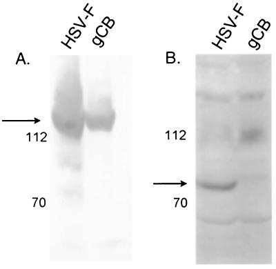
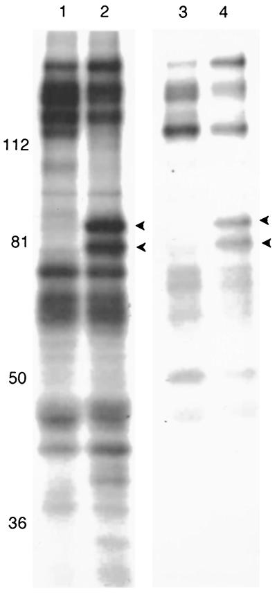
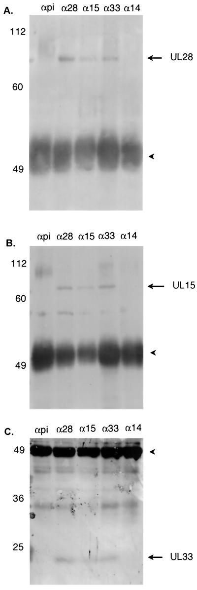
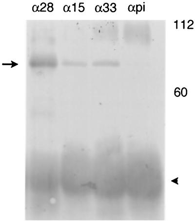
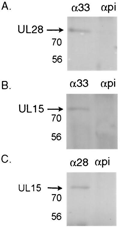
Previous studies have indicated that the U(L)6, U(L)15, U(L)17, U(L)28, U(L)32, and U(L)33 genes are required for the cleavage and packaging of herpes simplex viral DNA. To identify proteins that interact with the U(L)28-encoded DNA binding protein of herpes simplex virus type 1 (HSV-1), a previously undescribed rabbit polyclonal antibody directed against the U(L)28 protein fused to glutathione S-transferase was used to immunopurify U(L)28 and the proteins with which it associated. It was found that the antibody specifically coimmunoprecipitated proteins encoded by the genes U(L)28, U(L)15, and U(L)33 from lysates of both HEp-2 cells infected with HSV-1(F) and insect cells infected with recombinant baculoviruses expressing these three proteins. In reciprocal reactions, antibodies directed against the U(L)15- or U(L)33-encoded proteins also coimmunoprecipitated the U(L)28 protein. The coimmunoprecipitation of the three proteins from HSV-infected cells confirms earlier reports of an association between the U(L)28 and U(L)15 proteins and represents the first evidence of the involvement of the U(L)33 protein in this complex.


Figures

FIG. 1.
FIG. 1.
Digitally scanned images of immunoblots probed with anti-ICP8 antibody (A) and anti-UL28 antibody (B). Cells were infected with either HSV-1(F) or the UL28 mutant virus gCB, and the proteins were separated on an 8% polyacrylamide gel before being transferred to a nitrocellulose membrane and probed with antiserum. Protein sizes are indicated on the left in thousands.
FIG. 2.
FIG. 2.
Scanned digital image of fluorograph of radiolabeled proteins electrophoretically separated on an 8% polyacrylamide gel. HEp-2 cells were infected with either HSV-1(F) (lanes 1 and 2) or the gE-mutant virus strain 7202 (lanes 3 and 4) and immunoprecipitated as described in Materials and Methods by using either preimmune sera (lanes 1 and 3) or anti-UL28 antibody (lanes 2 and 4). Protein sizes are indicated on the left in thousands. Arrowheads indicate proteins of 85,000 and 80,000 apparent Mr, which correspond to the UL28 and UL15 proteins, respectively.
FIG. 3.
FIG. 3.
Scanned digital images of immunoblots probed with antisera directed against the UL28 (A), UL15 (B), or UL33 (C) proteins. Cell lysates were immunoprecipitated with preimmune serum (αpi) or antibody directed against UL28 (α28), UL15 (α15), UL33 (α33), or UL14 (α14), as described in the text. The position and size of molecular weight standards are indicated on the left in thousands. The heavychain of the immunoprecipitating antibody can be seen as a dark band around 49,000 (arrowhead). (A and B) 8% polyacrylamide gels; (C) 15% polyacrylamide gel.
FIG. 4.
FIG. 4.
Digitally scanned image of an immunoblot showing the detection of the UL28/UL15/UL33 protein complex from baculovirus-infected Sf21 cells. Cell lysates were immunoprecipitated as described in Materials and Methods with antibody directed against the UL28 (α28), UL15 (α15), or UL33 (α33) proteins or preimmune serum (αpi). The position of the UL28 protein is indicated with an arrow. The heavy chain of the antibody can be seen as a dark band at approximately 50,000 (arrowhead). Molecular weight standards are marked at the right in thousands.
FIG. 5.
FIG. 5.
Digitally scanned images of Western blots showing the proteins immunoprecipitated from dual-infected Sf21 cells. Cells were infected with baculoviruses expressing UL28 and UL33 (A), UL15 and UL33 (B), or UL28 and UL15 (C) and processed as described in Materials and Methods. Proteins were immunoprecipitated with antibody directed against the UL33 protein (α33) or the UL28 protein (α28), or with preimmune serum (αpi). An equivalent amount of lysate was used in each lane. Membranes were blotted with antibody directed against the UL28 protein (A) or UL15 protein (B and C). Protein sizes are indicated on the left in thousands.

References

    1. Abbotts, A. P., V. G. Preston, M. Hughes, A. H. Patel, and N. D. Stow. 2000. Interaction of the herpes simplex virus type 1 packaging protein UL15 with full-length and deleted forms of the UL28 protein. J. Gen. Virol. 81:2999-3009. - PubMed
    1. Addison, C., F. J. Rixon, J. W. Palfreyman, M. O'Hara, and V. G. Preston. 1984. Characterisation of a herpes simplex virus type 1 mutant which has a temperature-sensitive defect in penetration of cells and assembly of capsids. Virology 138:246-259. - PubMed
    1. Addison, C., F. J. Rixon, and V. G. Preston. 1990. Herpes simplex virus type 1 UL28 gene product is important for the formation of mature capsids. J. Gen. Virol. 71:2377-2384. - PubMed
    1. Adelman, K., B. Salmon, and J. D. Baines. 2001. Herpes simplex virus DNA packaging sequences adopt novel structures that are specifically recognized by a component of the cleavage and packaging machinery. Proc. Natl. Acad. Sci. USA 98:3086-3091. - PMC - PubMed
    1. al-Kobaisi, M. F., F. J. Rixon, I. McDougall, and V. G. Preston. 1991. The herpes simplex virus UL33 gene product is required for the assembly of full capsids. Virology 180:380-388. - PubMed

Publication types

LinkOut - more resources