Comparative Study
. 2002 May;76(10):4855-65.
doi: 10.1128/jvi.76.10.4855-4865.2002.

Ebola virus VP40 drives the formation of virus-like filamentous particles along with GP

Affiliations
Comparative Study

Ebola virus VP40 drives the formation of virus-like filamentous particles along with GP

Takeshi Noda et al. J Virol. 2002 May.

Abstract

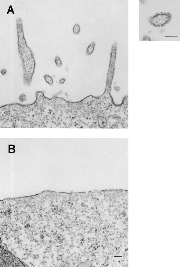
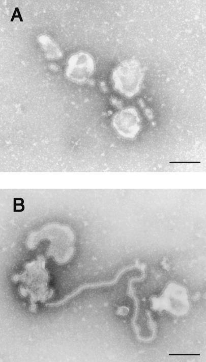
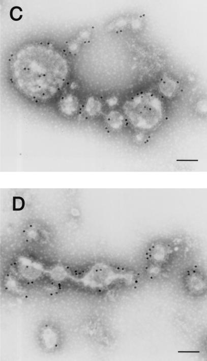
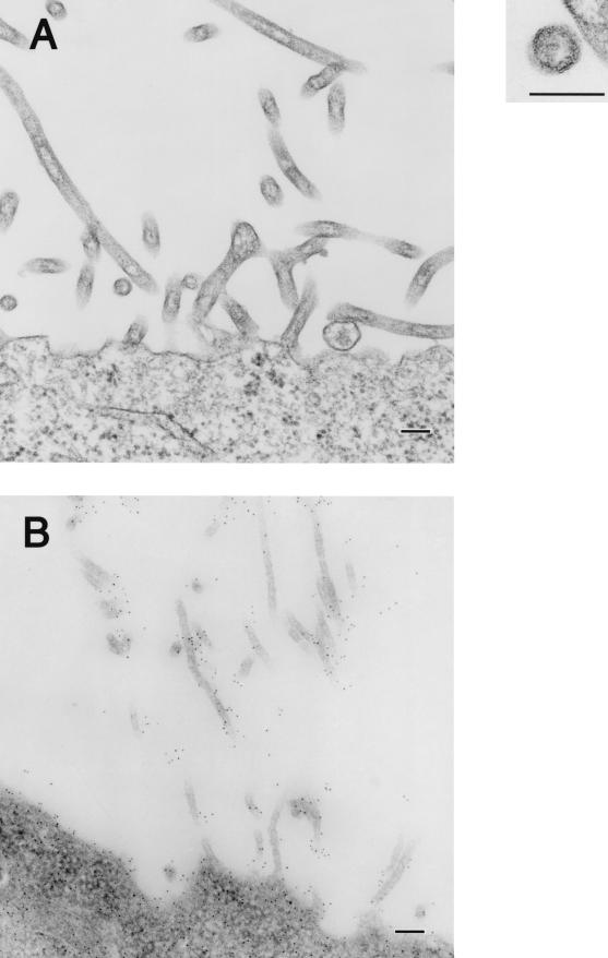
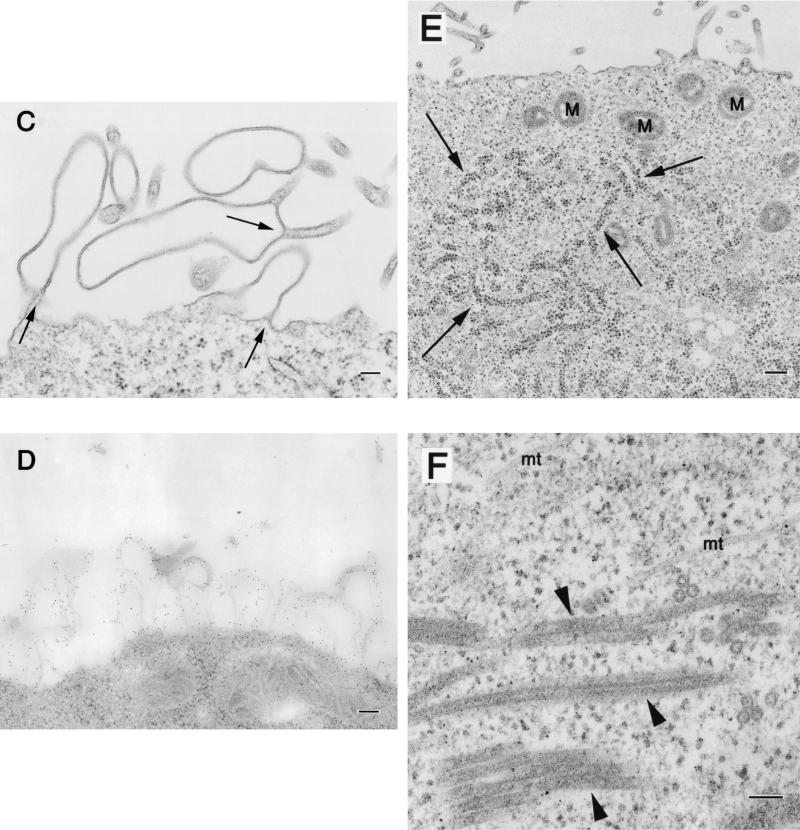
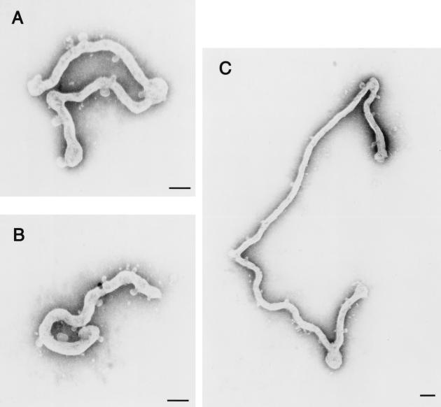
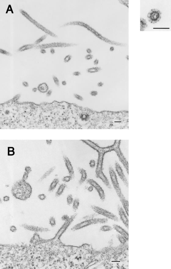
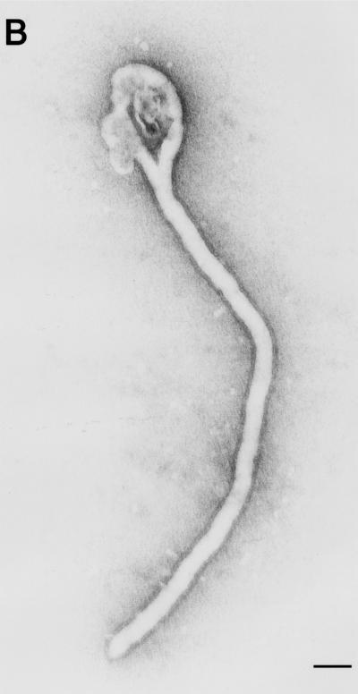
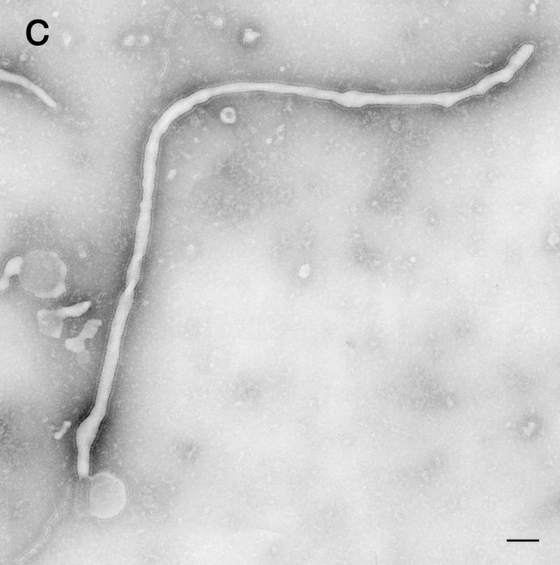
Using biochemical assays, it has been demonstrated that expression of Ebola virus VP40 alone in mammalian cells induced production of particles with a density similar to that of virions. To determine the morphological properties of these particles, cells expressing VP40 and the particles released from the cells were examined by electron microscopy. VP40 induced budding from the plasma membrane of filamentous particles, which differed in length but had uniform diameters of approximately 65 nm. When the Ebola virus glycoprotein (GP) responsible for receptor binding and membrane fusion was expressed in cells, we found pleomorphic particles budding from the plasma membrane. By contrast, when GP was coexpressed with VP40, GP was found on the filamentous particles induced by VP40. These results demonstrated the central role of VP40 in formation of the filamentous structure of Ebola virions and may suggest an interaction between VP40 and GP in morphogenesis.


Figures

FIG. 1.
FIG. 1.
Budding of GP-associated particles from the plasma membrane. (A) 293T cells at 24 h posttransfection with a GP-expressing plasmid. (B) 293T cells transfected with an empty expression vector lack such particle formation. Bars, 100 nm.
FIG. 2.
FIG. 2.
Pleomorphic particles resulting from GP expression. The supernatants of cells expressing GP were centrifuged through 20% sucrose, and the pelleted material was then negatively stained with 2% PTA. (A and B) Pleomorphic particles with surface spikes were observed. (C and D) Pelleted material was immunolabeled with a mixture of anti-GP monoclonal antibodies conjugated to 15-nm gold particles. Bars, 100 nm.
FIG. 2.
FIG. 2.
Pleomorphic particles resulting from GP expression. The supernatants of cells expressing GP were centrifuged through 20% sucrose, and the pelleted material was then negatively stained with 2% PTA. (A and B) Pleomorphic particles with surface spikes were observed. (C and D) Pelleted material was immunolabeled with a mixture of anti-GP monoclonal antibodies conjugated to 15-nm gold particles. Bars, 100 nm.
FIG. 3.
FIG. 3.
Morphological changes in 293T cells expressing VP40. At 24 h posttransfection of 293T cells with a VP40-expressing plasmid, filamentous particles budding from the plasma membrane (A), membrane ruffles and the adhering site of two bilayers (C, arrows), as well as aggregated ribosomes (E, arrows) were apparent. Intracellular electron-dense filamentous structures (F, arrowheads) were also observed. (B and D) The filamentous particles and membrane ruffles were immunolabeled with an anti-VP40 antibody conjugated with 5-nm gold particles. M, mitochondrion; mt, microtubule. Bars, 100 nm (panels A, B, C, D, and F) or 200 nm (panel E).
FIG. 3.
FIG. 3.
Morphological changes in 293T cells expressing VP40. At 24 h posttransfection of 293T cells with a VP40-expressing plasmid, filamentous particles budding from the plasma membrane (A), membrane ruffles and the adhering site of two bilayers (C, arrows), as well as aggregated ribosomes (E, arrows) were apparent. Intracellular electron-dense filamentous structures (F, arrowheads) were also observed. (B and D) The filamentous particles and membrane ruffles were immunolabeled with an anti-VP40 antibody conjugated with 5-nm gold particles. M, mitochondrion; mt, microtubule. Bars, 100 nm (panels A, B, C, D, and F) or 200 nm (panel E).
FIG. 4.
FIG. 4.
To determine how GP expression affects VP40-driven particle formation, we transfected 293T cells with both VP40- and GP-expressing plasmids. In ultrathin sections of the tranfected cells, we observed filamentous particle-like structures of 80-nm external diameter that were budding from the plasma membrane (Fig. 5).
FIG. 5.
FIG. 5.
Filamentous, spiked particles budding from the plasma membrane at 24 h posttransfection of 293T cells with plasmids coexpressing VP40 and GP. Bars, 100 nm.
FIG. 6.
FIG. 6.
Ebola virus-like particles produced by coexpression of VP40 and GP. The supernatants of cells coexpressing these two proteins were centrifuged through 20% sucrose, and the pelleted material was then negatively stained with 2% PTA. (A through C) Filamentous particles with surface spikes and varied lengths were observed. Pelleted material was immunolabeled with a mixture of anti-GP monoclonal antibodies conjugated to 15-nm gold particles (D, arrowheads), treated with 0.03% Triton X-100 at room temperature for 15 min, and then immunolabeled with a mixture of anti-GP antibodies conjugated to 15-nm gold particles (E, arrowheads) and an anti-VP40 antibody conjugated to 5-nm gold particles (E, arrows). Bars, 1 μm (panel A) or 100 nm (panels B through E).
FIG. 6.
FIG. 6.
Ebola virus-like particles produced by coexpression of VP40 and GP. The supernatants of cells coexpressing these two proteins were centrifuged through 20% sucrose, and the pelleted material was then negatively stained with 2% PTA. (A through C) Filamentous particles with surface spikes and varied lengths were observed. Pelleted material was immunolabeled with a mixture of anti-GP monoclonal antibodies conjugated to 15-nm gold particles (D, arrowheads), treated with 0.03% Triton X-100 at room temperature for 15 min, and then immunolabeled with a mixture of anti-GP antibodies conjugated to 15-nm gold particles (E, arrowheads) and an anti-VP40 antibody conjugated to 5-nm gold particles (E, arrows). Bars, 1 μm (panel A) or 100 nm (panels B through E).

References

    1. Campbell, S., and V. M. Vogt. 1997. In vitro assembly of virus-like particles with Rous sarcoma virus Gag deletion mutants: identification of the p10 domain as a morphological determinant in the formation of spherical particles. J. Virol. 71:4425-4435. - PMC - PubMed
    1. Coronel, E. C., K. G. Murti, T. Takimoto, and A. Portner. 1999. Human parainfluenza virus type 1 matrix and nucleoprotein genes transiently expressed in mammalian cells induce the release of virus-like particles containing nucleocapsid-like structures. J. Virol. 73:7035-7038. - PMC - PubMed
    1. Delchambre, M., D. Gheysen, D. Thines, C. Thiriart, E. Jacobs, E. Verdin, M. Horth, A. Burny, and F. Bex. 1989. The Gag precursor of simian immunodeficiency virus assembles into virus-like particles. EMBO J. 8:2653-2660. - PMC - PubMed
    1. Feldmann, H., and H. D. Klenk. 1996. Marburg and Ebola viruses. Adv. Virus Res. 47:1-52. - PubMed
    1. Gay, B., J. Tournier, N. Chazal, C. Carriere, and P. Boulanger. 1998. Morphopoietic determinants of HIV-1 Gag particles assembled in baculovirus-infected cells. Virology 247:160-169. - PubMed

Publication types