Comparative Study
. 2002 May;76(10):5062-70.
doi: 10.1128/jvi.76.10.5062-5070.2002.

Abnormal priming of CD4(+) T cells by dendritic cells expressing hepatitis C virus core and E1 proteins

Affiliations
Comparative Study

Abnormal priming of CD4(+) T cells by dendritic cells expressing hepatitis C virus core and E1 proteins

Pablo Sarobe et al. J Virol. 2002 May.

Erratum in

  • J Virol. 2004 Oct;138(1):193

Abstract

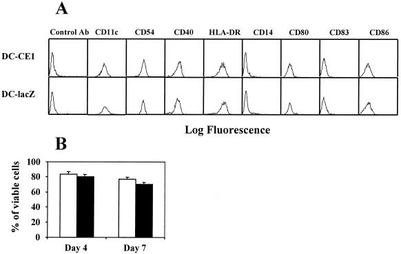
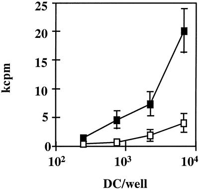
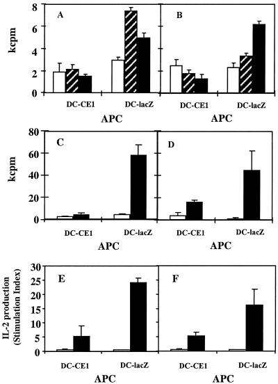
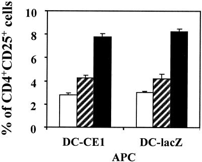
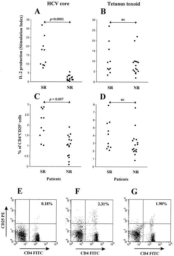
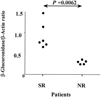
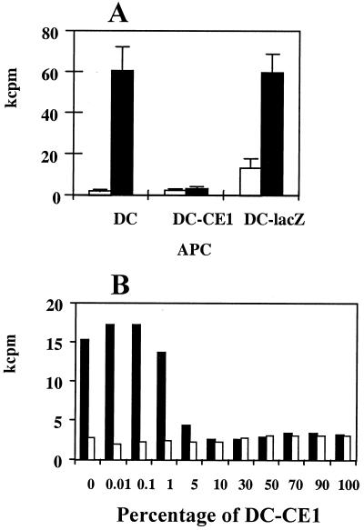
Patients infected with hepatitis C virus (HCV) have an impaired response against HCV antigens while keeping immune competence for other antigens. We hypothesized that expression of HCV proteins in infected dendritic cells (DC) might impair their antigen-presenting function, leading to a defective anti-HCV T-cell immunity. To test this hypothesis, DC from normal donors were transduced with an adenovirus coding for HCV core and E1 proteins and these cells (DC-CE1) were used to stimulate T lymphocytes. DC-CE1 were poor stimulators of allogeneic reactions and of autologous primary and secondary proliferative responses. Autologous T cells stimulated with DC-CE1 exhibited a pattern of incomplete activation characterized by enhanced CD25 expression but reduced interleukin 2 production. The same pattern of incomplete lymphocyte activation was observed in CD4(+) T cells responding to HCV core in patients with chronic HCV infection. However, CD4(+) response to HCV core was normal in patients who cleared HCV after alpha interferon therapy. Moreover, a normal CD4(+) response to tetanus toxoid was found in both chronic HCV carriers and patients who had eliminated the infection. Our results suggest that expression of HCV structural antigens in infected DC disturbs their antigen-presenting function, leading to incomplete activation of anti-HCV-specific T cells and chronicity of infection. However, presentation of unrelated antigens by noninfected DC would allow normal T-cell immunity to other pathogens.


Figures

FIG. 1.
FIG. 1.
Phenotypic analysis and cell viability of DC infected with recombinant adenovirus expressing HCV core and E1 genes or with control adenovirus. Monocyte-derived DC obtained from normal donors were cultured with cytokines for 7 days and infected with recombinant adenovirus (RAdCMVCE1 [DC-CE1] or RAdCMVLacZ [DC-lacZ]). (A) Twenty-four hours after infection cells were harvested and labeled with antibodies, and surface expressions of the different molecules were analyzed by flow cytometry. (B) RAdCMVCE1-infected DC (open bars) and RAdCMVLacZ-infected DC (filled bars) were cultured in the presence of purified T cells and stained with annexin V and propidium iodide 4 and 7 days after adenoviral infection. Results are expressed as the percentages of cells negative for both markers.
FIG. 2.
FIG. 2.
DC expressing HCV core and E1 genes have a lower allogeneic stimulatory capacity. Monocyte-derived DC obtained from normal donors were infected with RAdCMVCE1 (open squares) or with RAdCMVLacZ (filled squares). Twenty-four hours after infection, graded numbers of DC were incubated in flat-bottomed 96-well plates for 5 days with 105 nonadherent allogeneic cells obtained from a normal donor. [3H]thymidine was added for the last 18 h, and after harvesting, cell proliferation was measured in kilocounts per minute (kcpm). A representative case from four donors tested is shown.
FIG. 3.
FIG. 3.
DC expressing HCV core and E1 genes have poor stimulatory capacity for autologous primary and recall CD4+ T-cell responses. Monocyte-derived DC obtained from normal donors were infected with RAdCMVCE1 (DC-CE1) or with RAdCMVLacZ (DC-lacZ) and used as APC to stimulate CD4+ cells. DC (104) were incubated for 7 days with purified autologous CD4+ T cells (105) in the absence or in the presence of different antigens; proliferation was measured in kilocounts per minute (kcpm). (A and B) Induction of primary responses: cells were incubated without antigen (open bars) or in the presence of a 50 μM concentration of peptides p45 (hatched bars) or PADRE (filled bars). (C and D) Induction of recall responses: cells were incubated without antigen (open bars) or in the presence of 5 Lf of TT per ml (filled bars). Cells were pulsed with [3H]thymidine for the last 18 h of culture, and then they were harvested and proliferation was measured. (E and F) IL-2 production by CD4+ cells stimulated with DC incubated without antigen (open bars) or in the presence of 5 Lf of TT per ml (filled bars). Panels A to F show results from representative experiments obtained with six different donors.
FIG. 4.
FIG. 4.
CD25 expression of CD4+ T cells stimulated by DC. Monocyte-derived DC obtained from normal donors were infected with RAdCMVCE1 (DC-CE1) or with RAdCMVLacZ (DC-lacZ) and used to stimulate CD4+ T cells. DC were incubated with purified autologous CD4+ T cells in the absence (open bars) or in the presence of peptide PADRE (50 μM) (hatched bars) or TT (5 Lf/ml) (filled bars). Three days later, cells were double stained with phycoerythrin-labeled anti-CD25 and FITC-labeled anti-CD4 antibodies. Lymphocytes were gated, and 104 cells were analyzed for the expression of CD25 and CD4. Results represent the percentages of lymphocytes expressing both molecules. Results are representative of five different experiments.
FIG. 5.
FIG. 5.
IL-2 production and percentages of CD4+ CD25+ cells in response to HCV core or TT Antigens in HCV patients. PBMC from 10 SR patients and 16 NR patients were stimulated in vitro in the presence of 1 μg of HCV core antigen per ml (A and C) or 5 Lf of TT per ml (B and D). IL-2 production (expressed as SI) (A and B) and the percentages of CD4+ CD25+ cells as estimated by flow cytometry (C and D) were measured at days 7 and 3, respectively. Panels E to G are representative examples of the percentages of CD4+ CD25+ cells in a healthy seronegative control (E), an SR patient (F), and an NR patient (G) after stimulation with HCV core.
FIG. 6.
FIG. 6.
β-Glucuronidase mRNA expression in CD4+ CD25+ cells stimulated by HCV core in different groups of patients. CD4+ CD25+ cells were purified from six SR patients and five NR patients after 3 days of in vitro stimulation of PBMC in the presence of HCV core. mRNA was isolated, and reverse transcription-PCR for β-glucuronidase and β-actin was carried out. Resulting PCR bands were quantified, and the ratio of β-glucuronidase to β-actin for each patient is represented.
FIG. 7.
FIG. 7.
DC obtained from HCV patients maintain a good capacity to stimulate recall CD4+ T-cell responses. (A) Monocyte-derived DC obtained from patients with chronic hepatitis C were used to stimulate autologous CD4+ T cells incubated in the absence (open bars) or in the presence of 5 Lf of TT per ml (filled bars). Together with adenovirus-uninfected DC, DC infected with RAdCMVCE1 (DC-CE1) and with RAdCMVLacZ (DC-lacZ) were included in the same experiment. Results show T-cell proliferation obtained after 7 days in a representative case from a group of four patients tested. (B) Normal donor-derived DC were infected with RAdCMVCE1 or with RAdCMVLacZ, pooled at different proportions (keeping a total number of 104 DC) and used to stimulate autologous CD4+ T cells. They were incubated in the absence (open bars) or in the presence of 5 Lf of TT per ml (filled bars) for 7 days, and cell proliferation was measured in kilocounts per minute (kcpm).

Similar articles

Cited by

References

    1. Alexander, J., J. Sidney, S. Southwood, J. Ruppert, C. Oseroff, A. Maewal, K. Snoke, H. M. Serra, R. T. Kubo, A. Sette, and H. M. Grey. 1994. Development of high potency universal DR-restricted helper epitopes by modification of high affinity DR-blocking peptides. Immunity 1:751-761. - PubMed
    1. Auffermann-Gretzinger, S., E. B. Keeffe, and S. Levy. 2001. Impaired dendritic cell maturation in patients with chronic, but not resolved, hepatitis C virus infection. Blood 97:3171-3176. - PubMed
    1. Bain, C., A. Fatmi, F. Zoulim, J. P. Zarski, C. Trepo, and G. Inchauspe. 2001. Impaired allostimulatory function of dendritic cells in chronic hepatitis C infection. Gastroenterology 120:512-524. - PubMed
    1. Banchereau, J., and R. M. Steinman. 1998. Dendritic cells and the control of immunity. Nature 392:245-252. - PubMed
    1. Bruna-Romero, O., J. J. Lasarte, G. Wilkinson, K. Grace, B. Clarke, F. Borras-Cuesta, and J. Prieto. 1997. Induction of cytotoxic T-cell response against hepatitis C virus structural antigens using a defective recombinant adenovirus. Hepatology 25:470-477. - PubMed

Publication types

MeSH terms