. 2002 May;76(10):5121-30.
doi: 10.1128/jvi.76.10.5121-5130.2002.

Mutational analysis of simian virus 40 T-antigen primosome activities in viral DNA replication

Affiliations

Mutational analysis of simian virus 40 T-antigen primosome activities in viral DNA replication

Robert D Ott et al. J Virol. 2002 May.

Abstract

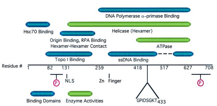
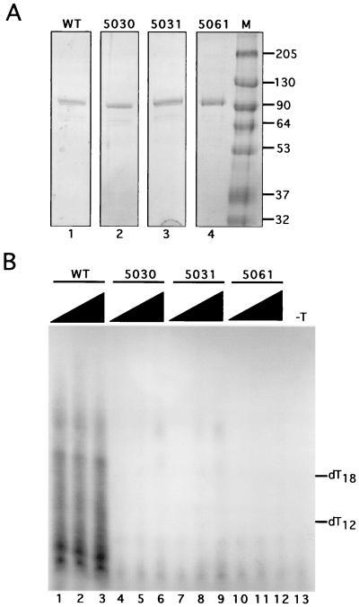
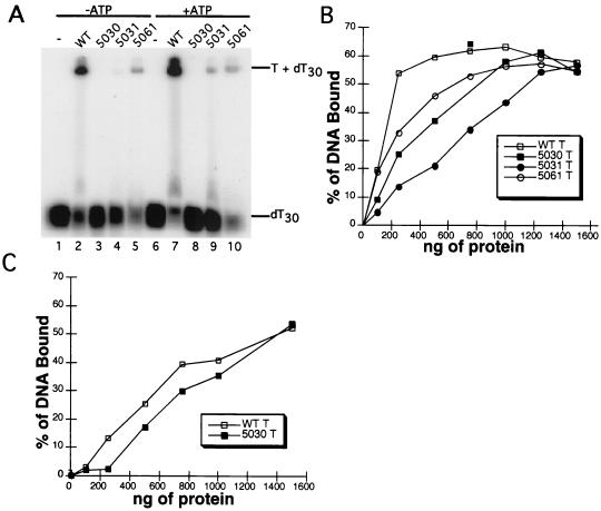
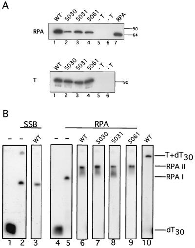
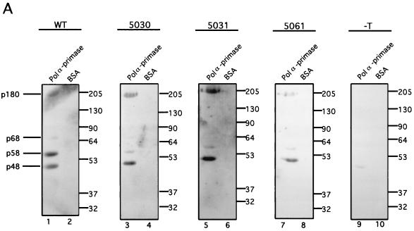
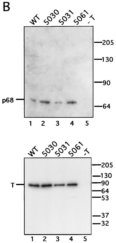
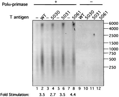
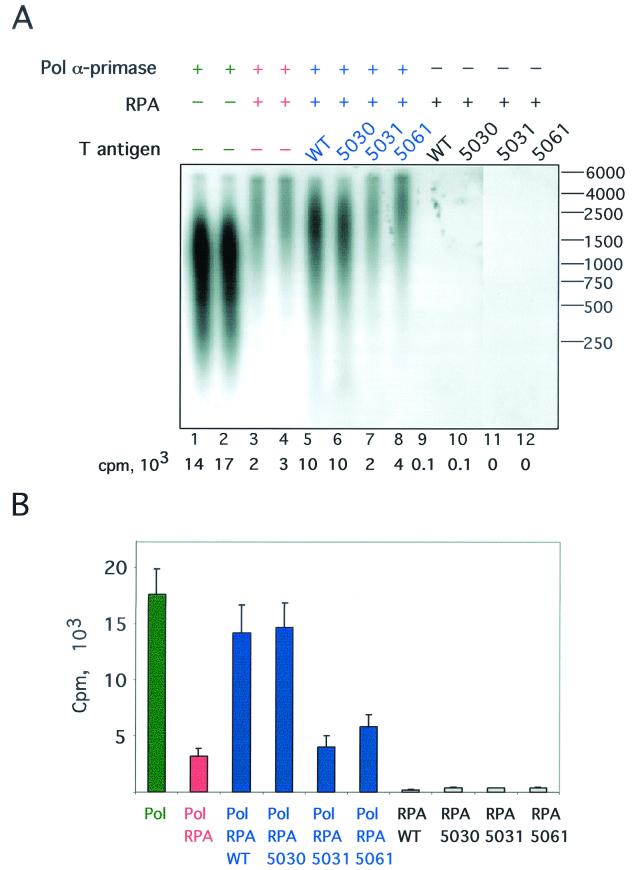
The recruitment of DNA polymerase alpha-primase (pol-prim) is a crucial step in the establishment of a functional replication complex in eukaryotic cells, but the mechanism of pol-prim loading and the composition of the eukaryotic primosome are poorly understood. In the model system for simian virus 40 (SV40) DNA replication in vitro, synthesis of RNA primers at the origin of replication requires only the viral tumor (T) antigen, replication protein A (RPA), pol-prim, and topoisomerase I. On RPA-coated single-stranded DNA (ssDNA), T antigen alone mediates priming by pol-prim, constituting a relatively simple primosome. T-antigen activities proposed to participate in its primosome function include DNA helicase and protein-protein interactions with RPA and pol-prim. To test the role of these activities of T antigen in mediating priming by pol-prim, three replication-defective T antigens with mutations in the ATPase or helicase domain have been characterized. All three mutant proteins interacted physically and functionally with RPA and pol-prim and bound ssDNA, and two of them displayed some helicase activity. However, only one of these, 5030, mediated primer synthesis and elongation by pol-prim on RPA-coated ssDNA. The results suggest that a novel activity, present in 5030 T antigen and absent in the other two mutants, is required for T-antigen primosome function.


Figures

FIG. 1.
FIG. 1.
Functional domains of SV40 T antigen in viral DNA replication. The protein regions necessary and sufficient for each of the indicated biochemical activities are diagrammed by the colored bars above the linear map of the amino acid sequence. Approximate domain boundaries inferred from partial proteolysis studies are indicated by the residue numbers below the linear sequence. Other features of the protein are depicted below the linear sequence. NLS, nuclear localization signal; circled P, clusters of phosphorylated Ser/Thr residues; Zn finger, structurally essential putative zinc-binding motif. The Walker A motif sequence ends at Thr-433. The genetic and biochemical mapping data summarized in the figure are published elsewhere (see references , , , , , , -, and and references therein).
FIG. 2.
FIG. 2.
Mutant SV40 T antigens fail to initiate SV40 DNA replication in the presence of purified initiation proteins. (A) Purified recombinant WT (lane 1), 5030 (lane 2), 5031 (lane 3), and 5061 (lane 4) T antigens were analyzed by SDS-10% PAGE and Coomassie staining. Lane M, marker proteins of known molecular masses (indicated in kilodaltons at the right). (B) Initiation of SV40 replication was assayed in reactions containing SV40 origin DNA, topoisomerase I, RPA, pol-prim, and 250, 500, or 1000 ng of WT or mutant SV40 large T antigen as indicated (lanes 1 to 12). Primers synthesized by pol-prim were visualized by denaturing 20% PAGE and autoradiography. Products synthesized in a control reaction without T antigen are shown (lane 13). The positions of end-labeled oligonucleotides dT12-18 used as markers are indicated.
FIG. 3.
FIG. 3.
ssDNA-binding activity of WT and mutant T antigens. (A) ssDNA-binding activity was tested in the presence (lanes 1 to 5) or absence of ATP (lanes 6 to 10). Reactions containing 10 pmol of WT (lanes 2 and 7), 5030 (lanes 3 and 8), 5031 (lanes 4 and 9) or 5061 (lanes 5 and 10) T antigen, as indicated, were incubated with end-labeled ssDNA in binding buffer for 30 min at 37°C. Protein-DNA complexes were visualized by native gel electrophoresis and autoradiography (overexposed here to show traces of bound DNA in lane 9). Lanes 1 and 6 show the products of a control reaction without T antigen. (B and C) Increasing amounts of T antigen were incubated with a partially duplex DNA template (B) or dT30 (C) for 20 min at 37°C. Protein-DNA complexes were immobilized on nitrocellulose filters, and bound DNA was quantitated by scintillation counting. The background was subtracted, and the percentage of input DNA bound was plotted as a function of input protein mass.
FIG. 4.
FIG. 4.
T-antigen interactions with RPA. (A) WT and the indicated mutant T antigens (3 μg of each) prebound to Pab101-Sepharose beads were incubated with purified RPA (5 μg) in a buffer analogous to that used in replication assays. Negative controls were carried out without T antigen. The beads were washed in the corresponding buffer, and immune complexes were resolved by SDS-PAGE. RPA was detected by immunoblotting with the monoclonal antibody 70C (upper panel). A sample of purified RPA run as a blotting control is shown in lane 7. The amount of T antigen in each reaction was verified by immunoblotting with the monoclonal antibody Pab101 (lower panel). The electrophoretic mobilities of marker proteins are noted at the right (in kilodaltons). (B) Reactions containing SSB-saturated ssDNA (lanes 2 and 3) or RPA-saturated ssDNA (lanes 5 to 9) were incubated with WT T antigen (lanes 3 and 6), 5030 (lane 7), 5031 (lane 8), and 5061 (lane 9) for 15 min at 37°C. Protein-DNA complexes were visualized by native gel electrophoresis and autoradiography. WT T antigen incubated with ssDNA in the absence of RPA formed a T-ssDNA complex as indicated (lane 10). Control reactions lacking protein are shown in lanes 1 and 4.
FIG. 5.
FIG. 5.
T-antigen binding to DNA polymerase α-primase. (A) WT and the indicated mutant T antigens were tested for the ability to interact with human pol-prim in a far-Western blot. First, 5 μg of purified pol-prim (lanes 1, 3, 5, 7, and 9) and an equivalent molar amount of BSA (lanes 2, 4, 6, 8, and 10) were resolved by SDS-PAGE and transferred to nitrocellulose. Then, the blots were incubated with purified T antigen at 10 μg/ml (lanes 1 to 8) or as a control without T antigen (lanes 9 to 10). Bound T antigen was detected with rabbit polyclonal anti-T antiserum and goat anti-rabbit horseradish peroxidase-conjugated secondary antibody. (The p180 band in lane 1 was partially obscured by a background smear of unknown origin.) The electrophoretic mobility of the pol-prim subunits is noted on the left, and the mobility of protein markers (in kilodaltons) is noted on the right. (B) A total of 3 μg of the indicated T antigen bound to Pab101-Sepharose beads was incubated with 4 μg of purified p68. The beads were washed, and immune complexes were resolved by SDS-PAGE (lanes 1 to 4). Control reactions were performed without T antigen (lane 5). The presence of p68 was detected by immunoblotting with the monoclonal antibody 9D5 (upper panel). The electrophoretic mobility of purified p68 is noted on the left, and the mobility of protein markers (in kilodaltons) is noted on the right. The amount of T antigen in each reaction was tested by immunoblotting with the monoclonal antibody Pab101 (lower panel).
FIG. 5.
FIG. 5.
T-antigen binding to DNA polymerase α-primase. (A) WT and the indicated mutant T antigens were tested for the ability to interact with human pol-prim in a far-Western blot. First, 5 μg of purified pol-prim (lanes 1, 3, 5, 7, and 9) and an equivalent molar amount of BSA (lanes 2, 4, 6, 8, and 10) were resolved by SDS-PAGE and transferred to nitrocellulose. Then, the blots were incubated with purified T antigen at 10 μg/ml (lanes 1 to 8) or as a control without T antigen (lanes 9 to 10). Bound T antigen was detected with rabbit polyclonal anti-T antiserum and goat anti-rabbit horseradish peroxidase-conjugated secondary antibody. (The p180 band in lane 1 was partially obscured by a background smear of unknown origin.) The electrophoretic mobility of the pol-prim subunits is noted on the left, and the mobility of protein markers (in kilodaltons) is noted on the right. (B) A total of 3 μg of the indicated T antigen bound to Pab101-Sepharose beads was incubated with 4 μg of purified p68. The beads were washed, and immune complexes were resolved by SDS-PAGE (lanes 1 to 4). Control reactions were performed without T antigen (lane 5). The presence of p68 was detected by immunoblotting with the monoclonal antibody 9D5 (upper panel). The electrophoretic mobility of purified p68 is noted on the left, and the mobility of protein markers (in kilodaltons) is noted on the right. The amount of T antigen in each reaction was tested by immunoblotting with the monoclonal antibody Pab101 (lower panel).
FIG. 6.
FIG. 6.
Stimulation of DNA polymerase α-primase by T antigen. Primer synthesis and elongation activity of limiting amounts of DNA pol-prim were assayed on ssDNA template in the absence (lanes 1, 3, 5, and 7) or the presence of 250 ng of WT (lane 2), 5030 (lane 4), 5031 (lane 6), and 5061 (lane 8) T antigens. Control reactions were carried out without pol-prim (lanes 9 to 12). Reaction products were resolved by alkaline electrophoresis, visualized by autoradiography, and quantitated by densitometry. The electrophoretic mobility of end-labeled marker DNA fragments of the indicated lengths in nucleotides is shown at the right.
FIG. 7.
FIG. 7.
Stimulation of DNA polymerase α-primase on RPA-coated ssDNA by T antigen. The ability of T antigen to stimulate primer synthesis and elongation by DNA pol-prim on RPA-coated ssDNA was tested. (A) Reactions were performed in duplicate and contained 300 ng of the indicated T antigen and 750 ng of RPA, pol-prim, ribo- and deoxyribonucleoside triphosphates, and radiolabeled dATP. The reaction products were resolved by alkaline gel electrophoresis and then visualized by autoradiography. Reaction products made in the absence of RPA (lanes 1 and 2) or pol-prim (lanes 9 to 12) are shown. The radioactivity (in counts per minute [cpm]) incorporated in each reaction is shown below each lane. The mobility of end-labeled marker DNA fragments of the indicated lengths in nucleotides is shown at the right. (B) Data from three independent experiments were quantitated, and the mean is plotted for each reaction. The brackets indicate the standard error of the mean.

Similar articles

Cited by

References

    1. Arezi, B., and R. D. Kuchta. 2000. Eukaryotic DNA primase. Trends Biochem. Sci. 25:572-576. - PubMed
    1. Barbaro, B. A., K. R. Sreekumar, D. R. Winters, A. E. Prack, and P. A. Bullock. 2000. Phosphorylation of simian virus 40 T-antigen on Thr124 selectively promotes double-hexamer formation on subfragments of the viral core origin. J. Virol. 74:8601-8613. - PMC - PubMed
    1. Beernink, H. T. H., and S. W. Morrical. 1999. RMPs: recombination/replication mediator proteins. Trends Biochem. Sci. 24:385-389. - PubMed
    1. Borowiec, J. A., F. B. Dean, and J. Hurwitz. 1991. Differential induction of structural changes in the simian virus 40 origin of replication by T antigen. J. Virol. 65:1228-1235. - PMC - PubMed
    1. Borowiec, J. A., F. B. Dean, P. A. Bullock, and J. Hurwitz. 1990. Binding and unwinding—how T antigen engages the SV40 origin of DNA replication. Cell 60:181-184. - PubMed

Publication types

MeSH terms

LinkOut - more resources