Comparative Study
. 2002 May;76(10):5167-83.
doi: 10.1128/jvi.76.10.5167-5183.2002.

Relationship between vaccinia virus intracellular cores, early mRNAs, and DNA replication sites

Affiliations
Comparative Study

Relationship between vaccinia virus intracellular cores, early mRNAs, and DNA replication sites

Massimo Mallardo et al. J Virol. 2002 May.

Abstract

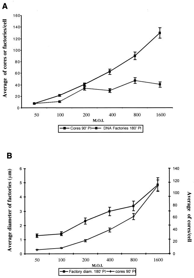
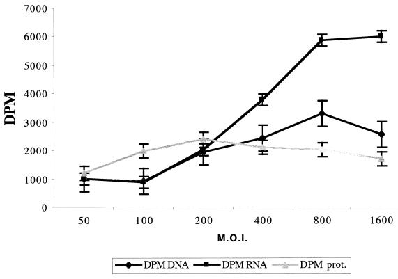
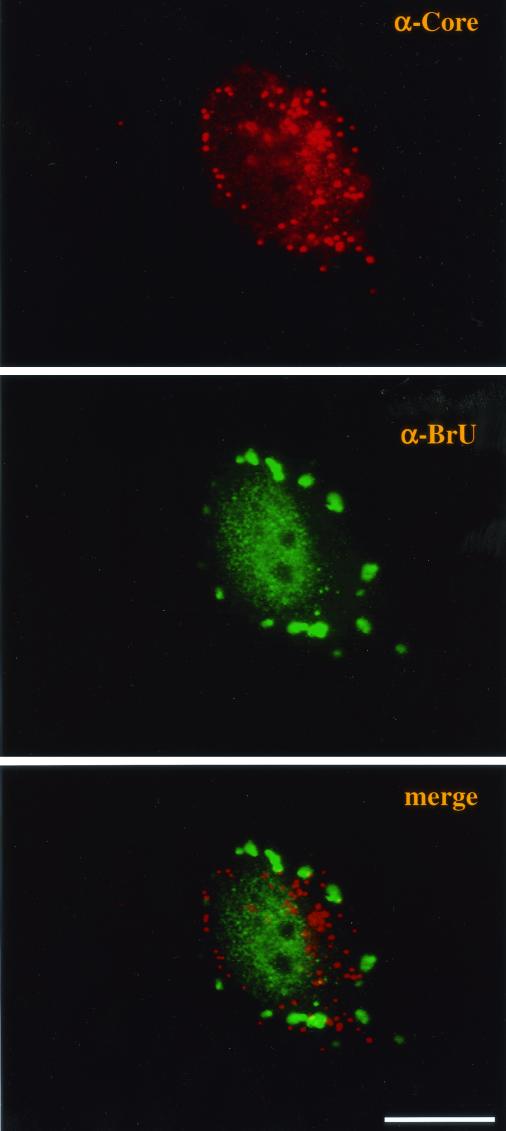
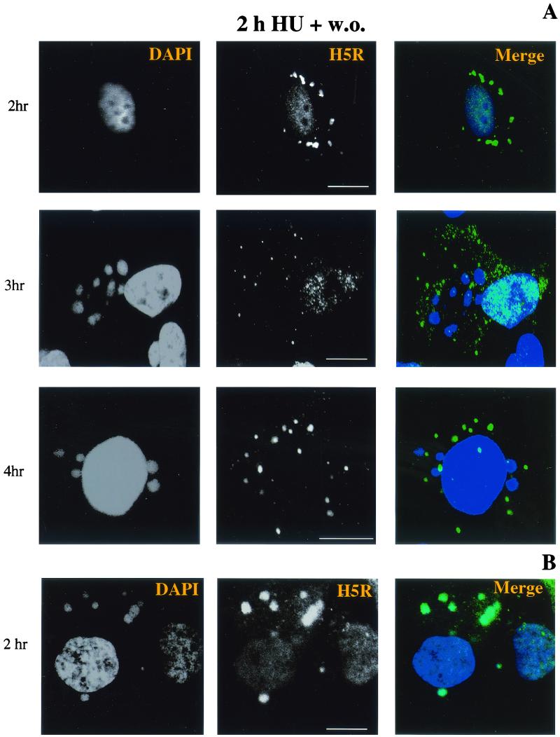
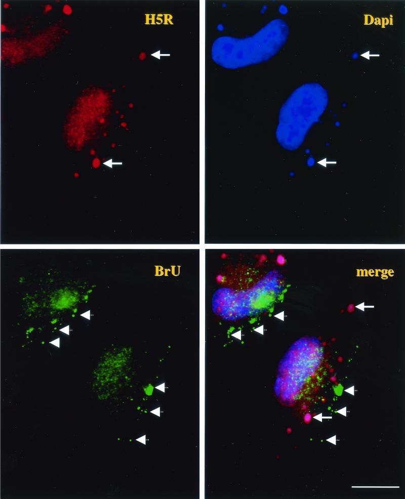
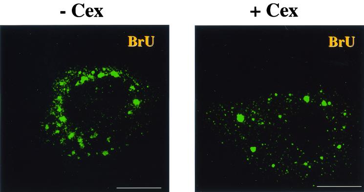
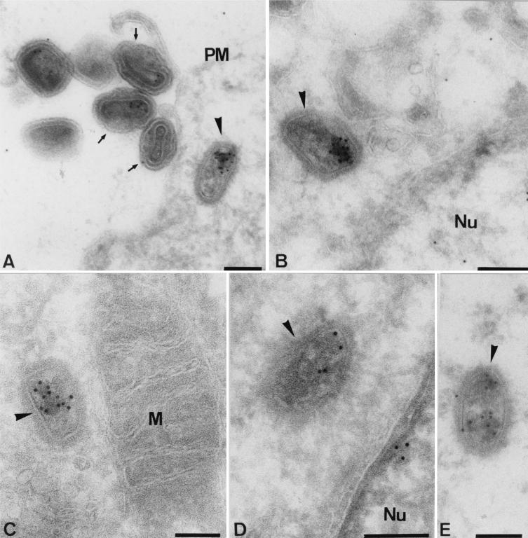
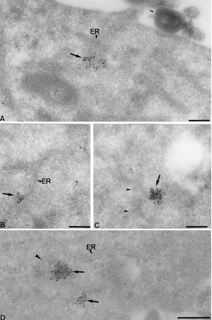
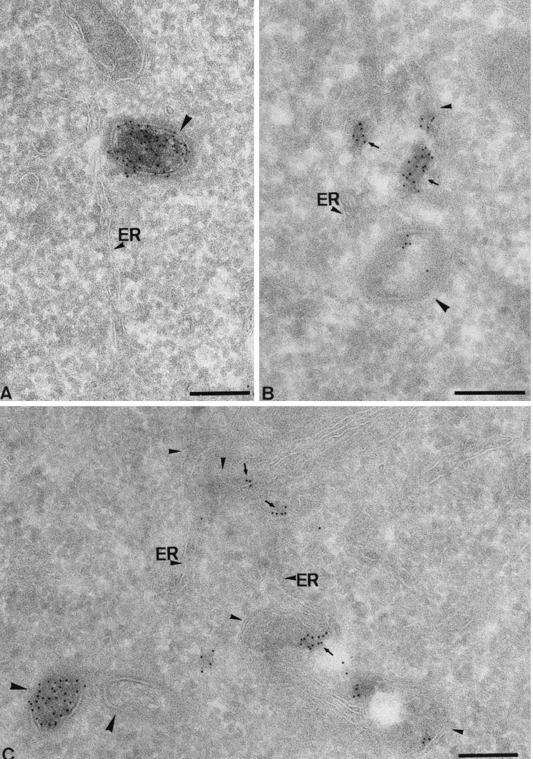
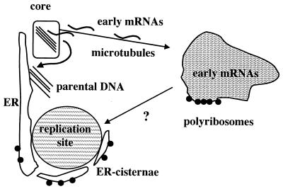
Virus assembly, a late event in the life cycle of vaccinia virus (VV), is preceded by a number of steps that all occur in the cytoplasm of the infected host cell: virion entry, delivery of the viral core into the cytoplasm, and transcription from these cores of early mRNAs, followed by the process of DNA replication. In the present study the quantitative and structural relationships between these distinct steps of VV morphogenesis were investigated. We show that viral RNA and DNA synthesis increases linearly with increasing amounts of incoming cores. Moreover, at multiplicities of infection that result in 10 to 40 cores per cell, an approximately 1:1 ratio between cores and sites of DNA replication exists, suggesting that each core is infectious. We have shown previously that VV early mRNAs collect in distinct granular structures that recruit components of the host cell translation machinery. Strikingly, these structures appeared to form some distance away from intracellular cores (M. Mallardo, S. Schleich, and J. Krijnse Locker, Mol. Biol. Cell 12:3875-3891, 2001). In the present study the intracellular locations of the sites of early mRNA accumulation and those of the subsequent process of DNA replication were compared. We show that these are distinct structures that have different intracellular locations. Finally, we study the fate of the parental DNA after core uncoating. By electron microscopy, cores were found close to membranes of the endoplasmic reticulum (ER) and the parental DNA, once it had left the core, appeared to associate preferentially with the cytosolic side of those membranes. Since we have previously shown that the process of DNA replication occurs in an ER-enclosed cytosolic "subcompartment" (N. Tolonen, L. Doglio, S. Schleich, and J. Krijnse Locker, Mol. Biol. Cell 12:2031-2046, 2001), the present data suggest that the parental DNA is released into the cytosol and associates with the same membranes where DNA replication is subsequently initiated. The combined data are discussed with respect to the cytosolic organization of VV morphogenesis.


Figures

FIG. 1.
FIG. 1.
Relation between MOI, the numbers of intracellular cores and sites of DNA replication, and the sizes of DNA replication sites. (A) Cells grown on coverslips were infected for 15 min at 37°C at the indicated MOI and fixed at 90 min postinfection (PI) in the presence of cycloheximide and 180 min postinfection without cycloheximide. Intracellular cores were counted at 90 min postinfection after labeling with the anti-core antibody, while replication sites were counted at 180 min after labeling with anti-p35. Values are average numbers ± standard errors of the means of cores or replication sites per cell in 30 cells and from three independent experiments. (B) Cells were infected at increasing MOIs, and intracellular cores counted in the same way as for panel A. The diameters of all the factories in 10 cells were measured with NIH Image at 180 min postinfection. Values are average diameters (in micrometers) ± standard errors of the means of all the factories in 10 cells.
FIG. 2.
FIG. 2.
Relation between MOI and the extents of early mRNA, DNA, and early protein synthesis. Cells were infected at the indicated MOIs as described in the legend to Fig. 1A and labeled either with [3H]uridine or [35S]methionine from 60 to 90 min postinfection or with [3H]thymidine from 150 to 180 min postinfection. After the labeling period, cells were lysed; equal amounts of OD595 units, measured by a Bio-Rad protein assay, were precipitated with trichloroacetic acid; and the amount of radioactivity contained in each sample was determined by liquid scintillation counting. Values are averages ± standard errors of the means of duplicate samples and from three independent experiments. Since the values for the [35S]methionine-labeled samples were much higher than those obtained for [3H]uridine or [3H]thymidine labeling, the [35S]methionine values in the graph represent the real values divided by 10.
FIG.3.
FIG.3.
Intracellular cores and the sites of early mRNA accumulation do not colocalize. HeLa cells grown on coverslips were infected and transfected with BrUTP as described in Materials and Methods in the presence of 5 mM HU in order to block DNA replication. Cells were fixed at 2 h postinfection and double labeled with the anti-core antibody, followed by donkey anti-rabbit coupled to rhodamine, and anti-BrU, followed by goat anti-rat-FITC. The merge in the bottom panel shows that the two structures do not overlap at all. Bar, 10 μm.
FIG.4.
FIG.4.
DNA replication may not be initiated where the early mRNAs accumulate. (A) Monolayers of cells grown on coverslips were infected and lipofected with a biotinylated antisense oligonucleotide corresponding to H5R mRNA as described in Materials and Methods in the presence of 2 mM HU. At 2 h postinfection, the HU was washed out and cells were incubated for an additional 1 and 2 h before being fixed and double labeled with DAPI and streptavidin-FITC (to detect the H5R oligonucleotide). The times postinfection are given on the left: 2 h postinfection corresponds to the time of HU washout, and 3 and 4 h postinfection correspond to 1 and 2 h post-HU washout, respectively. Note the absence of DAPI-positive replication sites at 2 h and the noncolocalization of the DAPI and H5R labels at 3 and 4 h. (B) Cells were infected for 2 h in the presence of HU, and the drug was then washed out, followed by transfection with the biotinylated H5R oligonucleotide. Cells were fixed at 2 h posttransfection (indicated on the left) and double labeled with DAPI and streptavidin-FITC (H5R). Note that under these conditions of infection and transfection, the DAPI- and streptavidin labels largely overlap. Bars, 10 μm.
FIG. 5.
FIG. 5.
Double transfection of BrUTP and the antisense H5R oligonucleotide at different times of infection reveals two different structures that accumulate viral mRNAs in infected cells. HeLa cells were infected and transfected with BrUTP for 2 h in the presence of 2 mM HU. HU was washed out, and cells were transfected with the antisense oligonucleotide corresponding to H5R mRNA. Double-transfected cells were fixed 2 h after HU washout and triple labeled with anti-BrU (FITC) (yellow channel), streptavidin-rhodamine (H5R) (red channel), and DAPI (bluechannel). The solid arrows in all four panels indicate DAPI-positive replication sites, the labeling of which entirely colocalizes with the pattern of the H5R oligonucleotide. The BrU pattern, indicated by hatched arrows (BrU and merge panels), does not colocalize with either of the two labels. It accumulates in granular structures that appear to have no relationship with the replication sites. This is most dramatically shown in the upper left cell, in which the replication sites all seem to collect at the upper left side of the cell, while the BrU-positive structures accumulate at the lower right side of the same cell. Bar, 10 μm.
FIG. 6.
FIG. 6.
The sites of mRNA accumulation develop in the same way in the presence of cycloheximide as in the absence of the inhibitor. HeLa cells were infected and transfected with BrUTP as described previously (19) in the absence (− Cex) or presence (+ Cex) of 5 μg of cycloheximide/ml. Infected cells were fixed at 2 h postinfection and labeled with anti-BrU. Bars, 10 μm.
FIG. 7.
FIG. 7.
Fate of the parental DNA under different conditions of infection. Monolayers of cells were infected at an MOI of 200 for 30 min at 37°C in the presence of either cycloheximide (A, B, and E), actD (C), or HU (D) and were fixed at 60 min (B and C) or 120 min (A, D, and E) postinfection. Cells were prepared for cryosectioning and labeled with anti-DNA. (A) Virions at the plasma membrane (PM) (small arrows) with low levels of label. One intracellular core (arrowhead) is clearly labeled. (B) A core close to the nucleus (Nu). (C) An intracellular core is found close to a mitochondrion (M). (D) The core accumulated in the presence of HU is less intensely labeled than the cores in the other images. (E) A core accumulated for 120 min in the presence of cycloheximide. Bars, 200 nm.
FIG. 8.
FIG. 8.
The fate of the parental DNA after core uncoating. (A through C) Cells were infected at an MOI of 200 in the presence of 5 mM HU and fixed at 2 h postinfection. Cryosections were labeled with anti-DNA. (A) An extracellular virion (small arrow) that is poorly labeled. In the cytoplasm one area (large arrow) can be seen that is heavily labeled and that seems associated with the ER (arrowhead). (B) The ER (indicated) seems to be labeled with anti-DNA at three positions, one of which is indicated by a large arrow. (C) A cytoplasmic labeled patch (arrow) is found close to ER membranes (arrowheads). (D) Cells infected for 1 h without HU. The image shows a core that is in the process of uncoating (large arrowhead) close to the ER (small arrowhead). DNA labeling is associated with the disassembling core as well as with the surrounding cytoplasm (large arrows). Bars, 200 nm.
FIG.9.
FIG.9.
Cores as well as the parental DNA lie close to ER membranes. All images are from cells infected for 1 h without inhibitor and labeled with anti-DNA. (A) A heavily labeled core (large arrowhead) that has apparently not yet uncoated its DNA can be seen close to the ER (small arrowhead). (B) A core (large arrowhead) weakly labeled with anti-DNA is found close to membranes of the ER (small arrowheads) that appear labeled (small arrows) on the cytosolic sides of the membranes. In this image the ER seems to bend around the DNA-containing structures. (C) Two cores can be seen (large arrowheads),of which one is unlabeled and one is heavily labeled. ER membranes are found close by (small arrowheads) and are labeled on their cytosolic sides (small arrows). In this image also, the ER membranes give the impression of bending around the DNA. Bars, 200 nm.
FIG. 10.
FIG. 10.
Model representing our view of the cytoplasmic organization of the early stages of VV infection. After entry, intracellular cores may associate with both MTs and the ER. From these cores early mRNAs are transcribed and extruded; they then organize in an MT-dependent fashion into typical (1-μm) granular structures. The latter are located some distance from the intracellular core and recruit polyribosomes as well as other components of the host cell translation machinery, making it likely that early proteins are synthesized at this site (19). Protein synthesis early in infection is required to induce the release of the parental DNA from the core. Upon release the parental DNA may associate with membranes of the ER. Early proteins involved in DNA synthesis are somehow recruited to the parental DNA (arrow with question mark) to initiate replication on the cytosolic side of the ER. As replication proceeds, new ER cisternae are recruited to the replication sites; these will eventually form an almost completely sealed ER envelope around the site (29). As the ER wrapping proceeds, membrane-bound ribosomes will preferentially be targeted to the outer ER membrane (black dots on the ER) (see reference 29).

Similar articles

Cited by

References

    1. Allan, V. 1996. Role of motor proteins in organizing the endoplasmic reticulum and Golgi apparatus. Semin. Cell Dev. Biol. 7:335-342.
    1. Beaud, G., and R. Beaud. 1997. Preferential virosomal location of underphosphorylated H5R protein synthesized in vaccinia virus-infected cells. J. Gen. Virol. 78:3297-3302. - PubMed
    1. Cairns, H. J. F. 1960. The initiation of vaccinia infection. Virology 11:603-623. - PubMed
    1. Cooper, J. A., and B. Moss. 1979. In vitro translation of immediate early, early, and late classes of RNA from vaccinia virus-infected cells. Virology 96:368-380. - PubMed
    1. Dahl, R., and J. R. Kates. 1970. Synthesis of vaccinia virus ‘early’ and ‘late’ messenger RNA in vitro with nucleoprotein structures isolated from infected cells. Virology 42:463-472. - PubMed

Publication types

LinkOut - more resources