. 2002 May;3(5):451-6.
doi: 10.1038/ni792. Epub 2002 Apr 22.

The CD40-TRAF6 axis controls affinity maturation and the generation of long-lived plasma cells

Affiliations

The CD40-TRAF6 axis controls affinity maturation and the generation of long-lived plasma cells

Cory Ahonen et al. Nat Immunol. 2002 May.

Abstract

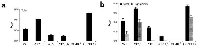
Affinity maturation of the immune response and the generation of long-lived bone marrow (BM) plasma cells are hallmarks of CD40-dependent, thymus-dependent (TD) humoral immunity. Through disruption of the tumor necrosis factor receptor (TNFR)-associated factor 6 (TRAF6)-binding site within the CD40 cytoplasmic domain, we selectively ablated affinity maturation and the generation of plasma cells after immunization. Mutagenesis of both the TRAF6 and TRAF2-TRAF3 sites was essential for arresting germinal center formation in response to immunization. CD40-induced B cell proliferation and early immunoglobulin production occurred even when all TRAF sites were ablated. These studies show that specific CD40-TRAF associations control well defined aspects of humoral immunity. In addition, they define the roles that TRAF-dependent and TRAF-independent pathways play in regulating antigen-driven B cell differentiation.


Conflict of interest statement

Competing interests statement: The authors declare that they have no competing financial interests.

Figures

Figure 1
Figure 1. Construction of X-CD40 transgenic mice
(a) The constructs used to generate X-CD40–transgenic mice consisted of the extracellular domain of human CD40 and the transmembrane and cytoplasmic domains of murine CD40. Amino acids targeted for conversion by site-directed mutagenesis are underlined. (b) X-CD40 expression on T cell–depleted splenocytes in each of the founder lines are shown. Filled histograms, anti–human CD40 staining of the transgenic receptor; open histograms, staining of an isotype-matched control antibody.
Figure 2
Figure 2. Signaling in X-CD40 transgenic mice relies on TRAF2/3 binding
(a) Primary cultures of splenic B cells from unimmunized X-CD40 transgenic mice were stimulated in vitro with 1000 ng/ml of shCD154 for various times. Cells were lysed and lysates were immunoblotted (IB) with antibodies to the phosphorylated TpY motif of Jnk2. Both isoforms of phosphorylated Jnk, p54 and p46, were recognized by the primary antibody. (b) Analysis of p38 MAPK activation in primary splenic B cells that were treated with shCD154. Cells were stimulated for various times, then lysed and immunoblotted with an antibody to the phosphorylated TpY motif of p38 MAPK. (c) Analysis of NF-κB activation in primary splenic B cells. Cells were treated with shCD154 for various times, lysed and immunoblotted with an antibody to Ser32-phosphorylated IκBα. For all signaling experiments, data are representative of three or more independent experiments. (d) Measurement of cellular IκBα content in B cells from transgenic mice. The nitrocellulose membrane in c was analyzed by immunoblotting with antibodies to IκBα, as a control for equivalent protein loading.
Figure 3
Figure 3. In vitro activation of B cells from X-CD40 mice
(a) Primary splenic B cells from each of the X-CD40 mice were stimulated in vitro with or without shCD154 (400 ng/ml) for 48 h. Cells were stained with biotinylated antibodies to CD23, which were detected with PE-streptavidin via flow cytometry. Data are the mean fluorescence intensity (MFI) of stained samples from cell cultures. Tg- denotes non-transgenic. (b) Proliferation was measured in splenic B cells treated with IL-4 + IL-5 either with or without shCD154 for 3 days. [3H]thymidine was added to cell cultures at 60 h. Cells were collected at 72 h and [3H]thymidine uptake was measured by scintillation counting. Data are mean±s.e.m. (c) IgM was measured in media samples taken from splenic B cells that were cultured for 5 days with IL-4 + IL-5 in the presence or absence of shCD154. IgM was measured by an isotype-specific ELISA. (d) Primary splenic B cells were cultured for 5 days with IL-4 + IL-5, in the presence or absence of shCD154. IgG1 was measured by an isotype-specific ELISA. All in vitro activation assays are representative of four or more experiments.
Figure 4
Figure 4. Humoral immune responses in X-CD40 mice
(a) Early IgG1 production was measured in vivo 15 days after intraperitoneal immunization with 100 μg of NP36-KLH that was emulsified in CFA. Circulating antibodies were measured by an NP-specific, isotype-specific ELISA. Data are mean±s.e.m. of three mice per group and are representative of three independent experiments. (b) Affinity maturation of the anti-NP response was measured 68 days after immunization with 100 μg of NP36-KLH emulsified in CFA. Circulating antibodies were quantified by an isotype-specific, NP-specific ELISA. NP25-BSA was used to capture all (total) antigen-specific Igs; NP4-BSA was used to capture only high-affinity Igs. Captured antibodies were detected with enzyme-conjugated anti-IgG1. Data are mean±s.e.m. of three mice per group and are representative of three independent experiments.
Figure 5
Figure 5. Generation of long-lived bone marrow plasma cells
(a) Mice were immunized with 100 μg of NP36-KLH emulsified in CFA. Mice were killed 68 days after immunization and bone marrow was collected. Bone marrow–derived lymphocytes were assessed for production of total antigen-specific IgG1 by ELISPOT; NP25-BSA was used as a capture antigen. (b) Generation of high-affinity plasma cells in the bone marrow of the same mice used in a. NP4-BSA was used as a capture antigen. Data are expressed as the number of antibody-secreting cells per 106 input cells and are representative of three or more independent experiments.
Figure 6
Figure 6. GC formation in the X-CD40 mice
(a) Immunohistological analysis of GC formation in spleens was done 10 days after immunization with sheep erythrocytes. Spleens were thinly sectioned, then stained with fluorochrome-coupled antibodies to CD4 and CD8 (red), PNA (blue) and CD19 (green). Images were captured by confocal microscope; they are representative of two experiments and >30 scanned images of three mice per group. (b) Flow cytofluorometric analysis of GC formation was done in mice immunized intraperitoneally with sheep erythrocytes and killed 10 days after immunization. Splenocytes were stained with fluorochrome-coupled antibodies to B220, PNA and GL7. Fluorescence was quantified by flow cytometry and profiles were gated on B220+ cells. Data are representative of three experiments with three mice per group.

References

    1. Banchereau J, et al. The CD40 antigen and its ligand. Annu Rev Immunol. 1994;12:881–922. - PubMed
    1. Kuhne MR, et al. Assembly and regulation of the CD40 receptor complex in human B cells. J Exp Med. 1997;186:337–342. - PMC - PubMed
    1. Pullen SS, et al. CD40-tumor necrosis factor receptor-associated factor (TRAF) interactions: regulation of CD40 signaling through multiple TRAF binding sites and TRAF hetero-oligomerization. Biochemistry. 1998;37:11836–11845. - PubMed
    1. Pullen SS, Dang TT, Crute JJ, Kehry MR. CD40 signaling through tumor necrosis factor receptor-associated factors (TRAFs). Binding site specificity and activation of downstream pathways by distinct TRAFs. J Biol Chem. 1999;274:14246–14254. - PubMed
    1. Cheng G, Baltimore D. TANK, a co-inducer with TRAF2 of TNF- and CD 40L-mediated NF-κB activation. Genes Dev. 1996;10:963–973. - PubMed

Publication types

MeSH terms