. 2002 Apr;14(4):931-43.
doi: 10.1105/tpc.010483.

Genes for two mitochondrial ribosomal proteins in flowering plants are derived from their chloroplast or cytosolic counterparts

Affiliations

Genes for two mitochondrial ribosomal proteins in flowering plants are derived from their chloroplast or cytosolic counterparts

Keith L Adams et al. Plant Cell. 2002 Apr.

Abstract

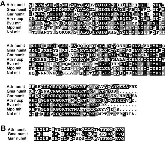
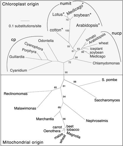
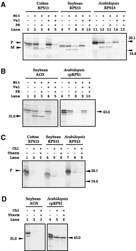
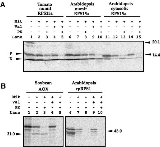
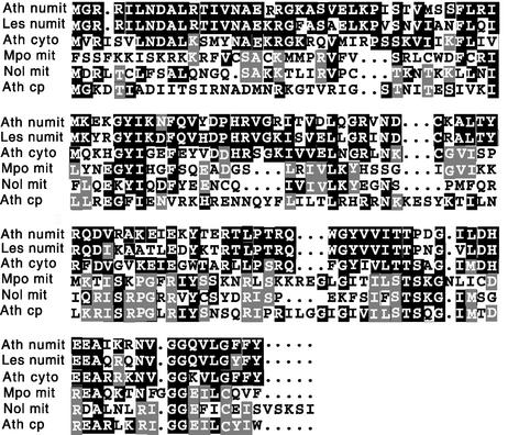
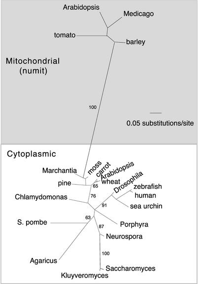
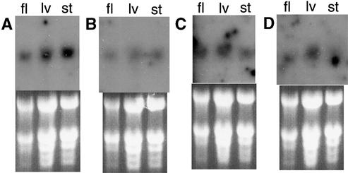
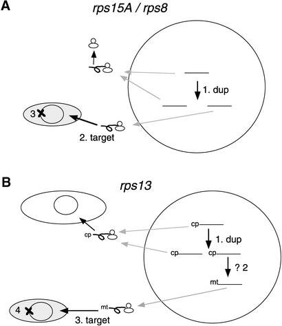
Often during flowering plant evolution, ribosomal protein genes have been lost from the mitochondrion and transferred to the nucleus. Here, we show that substitution by a duplicated, divergent gene originally encoding the chloroplast or cytosolic ribosomal protein counterpart accounts for two missing mitochondrial genes in diverse angiosperms. The rps13 gene is missing from the mitochondrial genome of many rosids, and a transferred copy of this gene is not evident in the nucleus of Arabidopsis, soybean, or cotton. Instead, these rosids contain a divergent nuclear copy of an rps13 gene of chloroplast origin. The product of this gene from all three rosids was shown to be imported into isolated mitochondria but not into chloroplasts. The rps8 gene is missing from the mitochondrion and nucleus of all angiosperms examined. A divergent copy of the gene encoding its cytosolic counterpart (rps15A) was identified in the nucleus of four angiosperms and one gymnosperm. The product of this gene from Arabidopsis and tomato was imported successfully into mitochondria. We infer that rps13 was lost from the mitochondrial genome and substituted by a duplicated nuclear gene of chloroplast origin early in rosid evolution, whereas rps8 loss and substitution by a gene of nuclear/cytosolic origin occurred much earlier, in a common ancestor of angiosperms and gymnosperms.


Figures

Figure 1.
Figure 1.
RPS13 Sequence Alignments. (A) Alignment of RPS13 sequences. Identical amino acids are shown on black and gray backgrounds. (B) Predicted targeting presequences of numit RPS13 sequences. Identical amino acids are shown in white on a black background. Dots indicate gaps inserted to improve alignment. Ath, Arabidopsis; Gma, Glycine max; Gar, Gossypium arboreum; Bvu, Beta vulgaris; Mpo, liverwort Marchantia polymorpha; Nol, green alga Nephroselmis olivacea.
Figure 2.
Figure 2.
Phylogenetic Analysis of Mitochondrial and Chloroplast rps13 Genes. Unrooted phylogram depicting the results of maximum likelihood analyses of the first and second nucleotide positions. The genes for numit rps13 from Arabidopsis, cotton, and legumes are indicated by asterisks; note the long branch lengths among these sequences compared with those for the nucp rps13 genes (genes for chloroplast rps13, located in the nucleus) from angiosperms. Genes located in the chloroplast genome are indicated by cp. The rps13 genes from the yeasts are located in the nucleus; other genes in the mitochondrial origin box are in the mitochondrial genome. Numbers indicate bootstrap values. See Methods for details of the analysis.
Figure 3.
Figure 3.
RPS13 Protein Import Experiments. (A) Import of RPS13 into isolated mitochondria. Lane 1, 35S-labeled precursor protein; lane 2, precursor protein incubated with soybean mitochondria; lane 3, as in lane 2 except that proteinase K was added after mitochondrial incubation to degrade unimported proteins; lanes 4 and 5, as in lanes 2 and 3, respectively, except that valinomycin was added before import to dissipate the membrane potential. Lanes 6 to 10 and 11 to 15 are equivalent to lanes 1 to 5. Markers are given in relative molecular mass (kD). Precursor (P) and mature (M) forms are indicated at left. (B) Import of the mitochondrial AOX protein (positive control) and the chloroplast protein RPS1 (cpRPS1; negative control) into isolated soybean mitochondria. Lanes are as in (A). (C) Import of RPS13 into isolated chloroplasts. Lane 1, 35S-labeled RPS13 precursor protein; lane 2, RPS13 precursor protein incubated with pea chloroplasts; lane 3, as in lane 2 except that thermolysin was added after chloroplast incubation to degrade unimported proteins. Lanes 4 to 6 and 7 to 9 are equivalent to lanes 1 to 3. (D) Import of the mitochondrial protein AOX (negative control) and the chloroplast protein RPS1 (positive control) into isolated pea chloroplasts. Lanes are as in (C). Chl, chloroplast; Mit, mitochondria; PK, proteinase K; Therm, thermolysin; Val, valinomycin; (+), presence; (−), absence.
Figure 6.
Figure 6.
Import of RPS15A into Mitochondria. (A) Import of RPS15A into soybean mitochondria. Lanes are the same as in Figure 3A. X denotes the ion front of the gel. (B) Import of the mitochondrial protein AOX and the chloroplast protein RPS1 (cpRPS1) into isolated soybean mitochondria. Lanes 1 to 5 and 6 to 10 are equivalent to lanes 1 to 5 of (A), except that the AOX and cpRPS1 precursor proteins were used. Mit, mitochondria; P, precursor; PK, proteinase K; Val, valinomycin.
Figure 4.
Figure 4.
Alignment of RPS15A and RPS8 Sequences. Identical amino acids are shown in white on a black or gray background. Dots indicate gaps inserted to improve alignment. The N-terminal extension of Marchantia RPS8 is not shown. Ath cp, chloroplast RPS8 from Arabidopsis; Ath cyto, cytosolic RPS15A from Arabidopsis; Ath numit, numit RPS15A from Arabidopsis; Les numit, numit RPS15A from tomato; Mpo mit, mitochondrial RPS8 from Marchantia; Nol mit, mitochondrial RPS8 from N. olivacea.
Figure 5.
Figure 5.
Phylogenetic Analysis of rps15A Genes. Unrooted phylogram of maximum likelihood analyses of the first and second nucleotide positions of an rps15A nucleotide alignment. Numbers indicate bootstrap values. See Methods for details of the analyses.
Figure 7.
Figure 7.
Expression Patterns of Four Nuclear Genes for Mitochondrial Ribosomal Proteins. (A) 3′ rpl2. (B) rps10. (C) Numit rps15A. (D) Numit rps13. Top panels show hybridizations, and bottom panels show the corresponding ethidium bromide–stained gels with prominent rRNA bands. The rps10 and 3′ rpl2 transcripts are ∼1.8 kb, the rps15A transcripts are ∼1.0 kb, and the rps13 transcripts have an approximate range of 900 to 1000 bp (with smaller transcripts present in flowers). fl, whole flowers; lv, rosette leaves; st, stems.
Figure 8.
Figure 8.
Models of Gene Substitution Events in the Evolution of Mitochondrial Ribosomal Proteins. Large circles indicate nuclei, gray ovals indicate mitochondria, and white ovals indicate chloroplasts. Black arrows indicate gene duplication (dup) or protein targeting to mitochondria (target); gray arrows indicate movement of gene transcripts to the cytosol. Bars indicate the mature coding regions of genes; targeting presequences are indicated by cp (chloroplast) and mt (mitochondrial). Proteins are indicated by loops attached to the ribosomes. Gene deletion (or pseudogene formation) is indicated by ×. (A) rps8/rps15A substitution. Step 1, cytosolic rps15A duplication; step 2, targeting of the duplicate to mitochondria; step 3, loss of rps8 from mitochondrial DNA. (B) rps13 substitution. Step 1, duplication of nucp rps13; step 2, possible acquisition of mitochondrial targeting presequence; step 3, targeting of the duplicate to mitochondria; step 4, loss of rps13 from mitochondrial DNA.

References

    1. Adams, K.L., Song, K., Roessler, P.G., Nugent, J.M., Doyle, J.L., Doyle, J.J., and Palmer, J.D. (1999). Intracellular gene transfer in action: Dual transcription and multiple silencings of nuclear and mitochondrial cox2 genes in legumes. Proc. Natl. Acad. Sci. USA 96, 13863–13868. - PMC - PubMed
    1. Adams, K.L., Daley, D.O., Qiu, Y.-L., Whelan, J., and Palmer, J.D. (2000). Repeated, recent and diverse transfers of a mitochondrial gene to the nucleus in flowering plants. Nature 408, 354–357. - PubMed
    1. Adams, K.L., Ong, H.C., and Palmer, J.D. (2001. a). Mitochondrial gene transfer in pieces: Fission of a ribosomal protein gene and partial or complete gene transfer to the nucleus. Mol. Biol. Evol. 18, 2289–2297. - PubMed
    1. Adams, K.L., Rosenblueth, M., Qiu, Y.-L., and Palmer, J.D. (2001. b). Multiple losses and transfers to the nucleus of two mitochondrial succinate dehydrogenase genes during angiosperm evolution. Genetics 158, 1289–1300. - PMC - PubMed
    1. Adams, K.L., Qiu, Y.-L., Stoutemyer, M., and Palmer, J.D. (2002). Punctuated evolution of mitochondrial gene content: High and variable rates of mitochondrial gene loss and transfer during angiosperm evolution. Proc. Natl. Acad. Sci. USA, in press. - PMC - PubMed

Publication types

Associated data