. 2002 May;22(10):3247-54.
doi: 10.1128/MCB.22.10.3247-3254.2002.

p53 Binds and activates the xeroderma pigmentosum DDB2 gene in humans but not mice

Affiliations

p53 Binds and activates the xeroderma pigmentosum DDB2 gene in humans but not mice

Thomas Tan et al. Mol Cell Biol. 2002 May.

Abstract

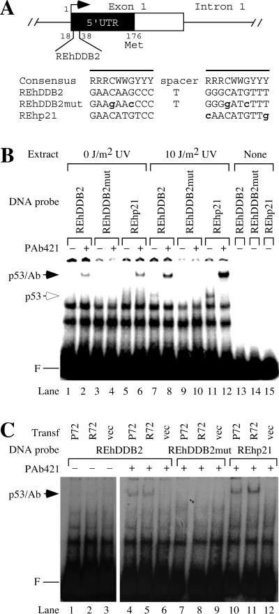
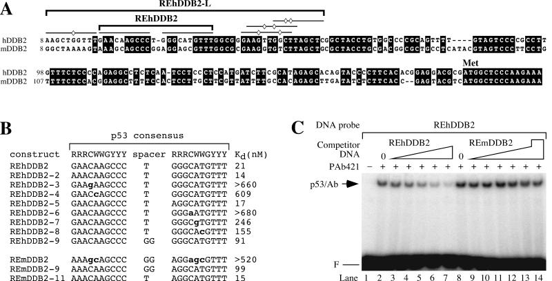
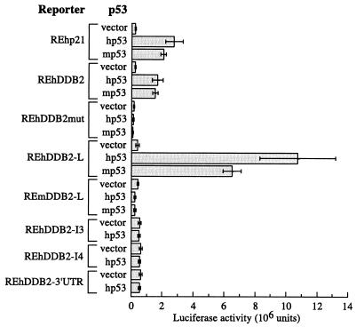
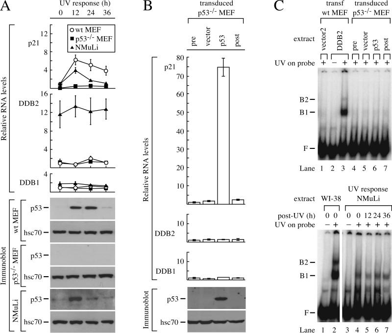
The DDB2 gene, which is mutated in xeroderma pigmentosum group E, enhances global genomic repair of cyclobutane pyrimidine dimers and suppresses UV-induced mutagenesis. Because DDB2 transcription increases after DNA damage in a p53-dependent manner, we searched for and found a region in the human DDB2 gene that binds and responds transcriptionally to p53. The corresponding region in the mouse DDB2 gene shared significant sequence identity with the human gene but was deficient for p53 binding and transcriptional activation. Furthermore, when mouse cells were exposed to UV, DDB2 transcription remained unchanged, despite the accumulation of p53 protein. These results demonstrate direct activation of the human DDB2 gene by p53. They also explain an important difference in DNA repair between humans and mice and show how mouse models can be improved to better reflect cancer susceptibility in humans.


Figures

FIG. 1.
FIG. 1.
The 5′ UTR of the human DDB2 gene contains a consensus sequence for p53 binding. (A) Consensus p53 binding site in the human DDB2 gene. The putative p53 response element from the human DDB2 gene (REhDDB2) is located from 18 to 38 bp downstream of the putative transcriptional start site. The consensus p53 binding site is shown, where R stands for purines A or G, Y stands for pyrimidines C or T, and W stands for A or T. The boldface lowercase letters indicate deviations from the consensus sequence. The lines denote the two half sites for p53 binding. REhDDB2mut was derived from REhDDB2 by mutating four highly conserved sites in the p53 consensus and served as a negative control in the experiments. REhp21 is the p53 response element from the human p21 gene and served as a positive control in the experiments. (B) Binding of p53 in HT1080 cell extracts to REhDDB2. Nuclear extracts were made from HT1080 cells, which had been untreated or treated with UV at a dose of 10 J/m2. A 32P-labeled DNA probe was incubated with the extracts and resolved by EMSA. F marks the position of the free DNA probe. Where indicated, incubations also included the monoclonal antibody PAb421, which binds to p53 and stabilizes its binding activity. The mobilities of the p53-DNA complex and the p53-PAb421-DNA complex are indicated (p53 and p53/Ab, respectively). (C) Binding of p53 in transfected cell extracts to REhDDB2. Nuclear extracts were made from 041 mut (p53−/−) cells, which had been transfected with vector or with one of the common wild-type alleles of p53, P72 or R72. Different 32P-labeled DNA probes were incubated with extracts with or without PAb421 and resolved by EMSA.
FIG. 2.
FIG. 2.
The 5′ UTRs in the human and mouse DDB2 genes share high sequence identity and contain several consensus p53 binding sites. (A) Alignment of the human and mouse DDB2 genes. The lines indicate putative p53 binding half sites. The diamonds indicate deviations from the consensus p53 binding site for the human DDB2 gene. The bracketed REhDDB2 was confirmed as a p53 binding site in Fig. 1 (Kd = 21 nM). The bracketed REhDDB2-L is a larger region of the human DDB2 gene confirmed as an even better p53 binding site (Kd = 4 nM). (B) Dissociation constants for p53 binding to different response element constructs. Various DNA constructs derived from the consensus p53 response elements from the human and mouse DDB2 genes were tested for their abilities to compete for binding to in vitro-translated p53 in a competition EMSA such as that shown in panel C. The Kd for REhDDB2 was determined from the fit to equation 2, as described in Materials and Methods. All other dissociation constants were determined from the fit to equation 1. Competition was not observed to any significant degree for REhDDB2-3, REhDDB2-6, or REmDDB2. Therefore, the Kd shown is greater than the highest concentration of cold competitor tested. The boldface lowercase letters indicate deviations from the consensus p53 binding site. R, Y, and W are defined as in the Fig. 1A legend. (C) Competition EMSA for p53 binding. In vitro-translated p53 was incubated with PAb421 and 32P-labeled REhDDB2 DNA probe (0.3 nM) together with unlabeled competitor DNA and resolved by EMSA. Competitor DNA was either REhDDB2 or REmDDB2 at a concentration of 12.5 nM (lanes 3 and 9), 25 nM (lanes 4 and 10), 50 nM (lanes 5 and 11), 100 nM (lanes 6 and 12), or 200 nM (lanes 7 and 13) and was resolved by EMSA. For REmDDB2, a concentration of 520 nM was also tested in lane 14. F and p53/Ab are defined as in the Fig. 1 legend.
FIG. 3.
FIG. 3.
p53 activates reporters containing REhDDB2 and REhDDB2-L but not REmDDB2-L, REhDDB2-I3, REhDDB2-I4, or REhDDB2-3′ UTR. Luciferase reporters containing various DNA elements were cotransfected with human p53 (hp53), mouse p53 (mp53), or vector alone into 041 mut (p53−/−) cells. Extracts from the transfections were assessed for luciferase activity. The bars represent means of the results of two to seven independent experiments, each done in sextuplicate.
FIG. 4.
FIG. 4.
Transcription of DDB2 does not respond to p53 in mouse cells. (A) Response of mouse cells to UV. Wild-type MEF, mutant p53−/− MEF, and a normal mouse liver cell line (NMuLi) were exposed to 10 J of UV/m2 and harvested for protein and cytoplasmic RNA after 12, 24, and 36 h. Quantitative RT-PCR was performed for p21, DDB2, and DDB1, and the RNA levels for each gene were normalized to the 0-h time point of wild-type MEF. Immunoblots were performed for p53 and hsc70. (B) Response of p53−/− MEF to transduction of p53. p53−/− MEF were transduced with virus expressing mouse p53 or vector and harvested for protein and cytoplasmic RNA after 48 h. Untransduced cells were also harvested at time points 0 (pre) and 48 (post) h. Quantitative RT-PCR was performed for p21, DDB2, and DDB1, and the RNA levels were normalized to pretransduction levels. Immunoblots were performed for p53 and hsc70. (C) UV-DDB after p53 transduction or UV exposure. Cell extracts were incubated with a 148-bp 32P-labeled DNA probe that was nonirradiated (−) or irradiated with 5,000 J of UV/m2 (+). The upper panel shows an EMSA for UV-DDB in extracts from p53−/− MEF transduced with empty vector (lane 5) or mouse p53 (lane 6). Also included are controls with extracts from untransduced p53−/− MEF at 0 (pre) and 48 (post) h (lanes 4 and 7, respectively) and extracts from wild-type MEF transfected with vector2 (lane 1) or mouse DDB2 (lanes 2 and 3). The lower panel shows an EMSA for UV-DDB in NMuLi extracts 12, 24, and 36 h after UV exposure. Extracts from human wild-type fibroblasts (WI-38; lanes 1 and 2) were included to show the higher levels of UV-DDB in human cells compared to NMuLi. F is defined as in the Fig. 1 legend. wt, wild type; B1 and B2, complexes of UV-damaged DNA bound to UV-DDB.

References

    1. Andrews, N. C., and D. V. Faller. 1991. A rapid micropreparation technique for extraction of DNA-binding proteins from limiting numbers of mammalian cells. Nucleic Acids Res. 19:2499.. - PMC - PubMed
    1. Beckman, G., R. Birgander, A. Sjalander, N. Saha, P. A. Holmberg, A. Kivela, and L. Beckman. 1994. Is p53 polymorphism maintained by natural selection? Hum. Hered. 44:266-270. - PubMed
    1. Bissonnette, N., and D. J. Hunting. 1998. p21-induced cycle arrest in G1 protects cells from apoptosis induced by UV-irradiation or RNA polymerase II blockage. Oncogene 16:3461-3469. - PubMed
    1. Bohr, V., C. Smith, D. Okumoto, and P. Hanawalt. 1985. DNA repair in an active gene: removal of pyrimidine dimers from the DHFR gene of CHO cells is much more efficient than in the genome overall. Cell 40:359-369. - PubMed
    1. Chu, G., and E. Chang. 1988. Xeroderma pigmentosum group E cells lack a nuclear factor that binds to damaged DNA. Science 242:564-567. - PubMed

Publication types