. 2002 Apr 30;99(9):5971-6.
doi: 10.1073/pnas.052159099. Epub 2002 Apr 23.

Requirement of Ca2+ and CaMKII for Stat1 Ser-727 phosphorylation in response to IFN-gamma

Affiliations

Requirement of Ca2+ and CaMKII for Stat1 Ser-727 phosphorylation in response to IFN-gamma

Jayasree S Nair et al. Proc Natl Acad Sci U S A. .

Abstract

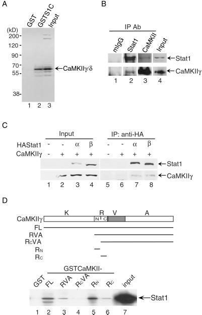
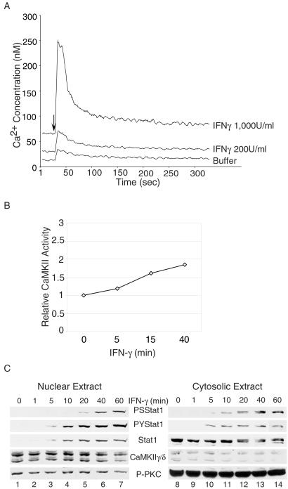
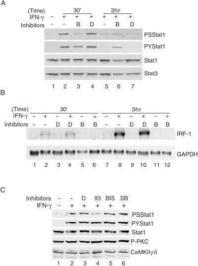
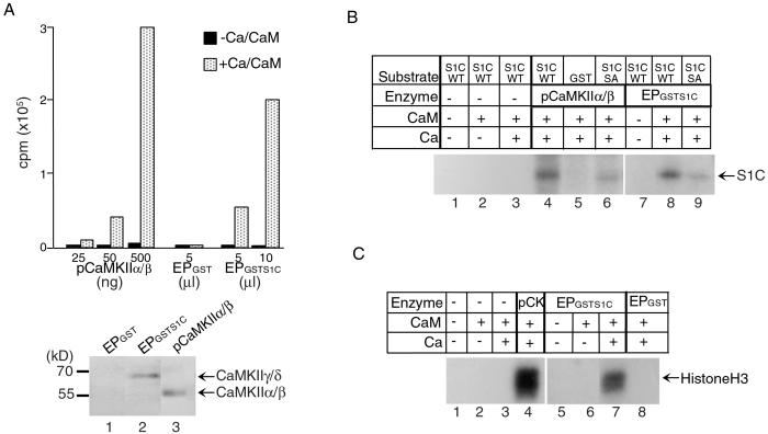
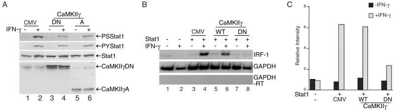
In response to IFN-gamma, the latent cytoplasmic protein signal transducers and activators of transcription 1 (Stat1) becomes phosphorylated on Y701, dimerizes, and accumulates in the nucleus to activate transcription of IFN-gamma-responsive genes. For maximal gene activation, S727 in the transcription activation domain of Stat1 also is inducibly phosphorylated by IFN-gamma. We previously purified a group of nuclear proteins that interact specifically with the Stat1 transcription activation domain. In this report, we identified one of them as the multifunctional Ca(2+)/calmodulin-dependent kinase (CaMK) II. We demonstrate that IFN-gamma mobilizes a Ca(2+) flux in cells and activates CaMKII. CaMKII can interact directly with Stat1 and phosphorylate Stat1 on S727 in vitro. Inhibition of Ca(2+) flux or CaMKII results in a lack of S727 phosphorylation and Stat1-dependent gene activation, suggesting in vivo phosphorylation of Stat1 S727 by CaMKII. Thus two different cellular signaling events, IFN-gamma receptor occupation and Ca(2+) flux, are required for Stat1 to achieve maximal transcriptional activation through regulation of phosphorylation.


Figures

Figure 1
Figure 1
Specific interactions between Stat1 and CaMKII in vivo and in vitro. (A) GST pull-down assays were performed with GST-fusion proteins containing the Stat1 TAD and nuclear extract of U3A cells. The bound proteins were analyzed by Western blotting. S1C, Stat1 TAD. (B) Coimmunoprecipitation (IP) analyses were performed with whole-cell extracts from NIH 3T3 cells. The endogenous Stat1 or CaMKII proteins in the immune complexes were analyzed by Western blotting. mIgG, control antibody from mouse serum. (C) Whole-cell extracts were prepared from U3A cells transfected with CaMKII-γ and HA-tagged Stat1-α or -β. The immune-precipitated complexes by anti-HA were analyzed by Western blotting. (D) GST pull-down assays were performed with GST-fusion proteins containing the various portions of CaMKIIγ and in vitro translated 35S-labeled Stat1. The bound proteins were separated by SDS/PAGE and visualized by autoradiography. K, kinase domain (residues 1–272); R, regulatory domain (273–315); V, variable region (316–385); A, association domain (386–518); Rn, amino-terminal half of the regulatory domain (273–290); Rc, carboxyl-terminal half of the regulatory domain (291–315); FL, full length (1–518).
Figure 2
Figure 2
IFN-γ induces Ca2+ transients and activates CaMKII. (A) NIH 3T3 cells were preloaded with fluo-3 AM, and the [Ca2+]i was measured at 1-sec intervals after IFN-γ treatment by a Fluroskan Ascent FL fluorospectrophotometer (Labsystems) and analyzed by the ASCENT 2.4 software (Labsystems). The arrow indicates the addition of IFN-γ to the cells. (B) NIH 3T3 cells were treated with 200 units/ml mouse IFN-γ for the times indicated, and cell lysates were analyzed in a CaMKII kinase assay with the CaMKII-selective autocamtide-3 as substrate. (C) Nuclear and cytoplasmic extracts were prepared from NIH 3T3 cells treated with 200 units/ml mouse IFN-γ for the times indicated and analyzed by Western blotting. PS, Phospho-S727; PY, Phospho-Y701; P-PKC, activated phospho-PKC.
Figure 3
Figure 3
Requirement of Ca2+ and CaMKII for IFN-γ-induced phosphorylation of Stat1 S727 and gene activation. (A) Whole-cell extracts were prepared from NIH 3T3 cells with the indicated treatment and analyzed by Western blotting. Pretreatments with DMSO (D) or 50 μM BAPTA-AM (B) were for 30 min followed by 200 units/ml mouse IFN-γ for the length of time indicated. PS, phospho-S727; PY, phospho-Y701. (B) Total RNAs were prepared from NIH 3T3 cells with the indicated treatment as described for A and analyzed by Northern blotting. (C) NIH 3T3 cells were pretreated with the indicated reagents for 30 min followed by 200 units/ml mouse IFN-γ for 30 min, and whole-cell extracts were analyzed by Western blotting. 93 , KN93 (5 μM); BIS, bisindolylmaleimide 1 (20 μM); SB, SB203580 (10 μM); P-PKC, activated phospho-PKC.
Figure 4
Figure 4
CaMKII phosphorylates Stat1 S727 in vitro. (A Upper) Kinase assays were performed with purified rat brain CaMKIIα/β and a CaMKIIγ/δ-containing fraction purified from U3A nuclear extracts by using GST-Stat1 TAD affinity columns (EPGSTS1C) or eluates from a GST column (EPGST) as control. The incorporation of 32P in the CaMKII substrate, autocamtide-3, was measured by a scintillation counter. (Lower) Western blot analyses of 10 μl of eluates from a GST column (lane 1), from a GST-Stat1 TAD affinity column (lane 2), and 250 ng of pCaMKIIα/β (lane 3). S1C, Stat1 TAD; pCaMKII, purified CaMKIIα/β, Ca, calcium; CaM, calmodulin; EP, eluted protein. (B) GST-fusion proteins containing WT or S727A mutant Stat1 TAD were used as substrates for the indicated CaMKIIs. Approximately 250 ng of CaMKIIα/β, 200 ng of CaMKIIγ/δ, and 2.5 μg of various GST-fusion proteins were used in the kinase assays. The incorporation of 32P in the Stat1 TAD was visualized by autoradiography after SDS/PAGE. SA, S727A. (C) Purified histone H3 (1 μg) was used as a substrate for the indicated CaMKIIs, and the incorporation of 32P was visualized by autoradiography after SDS/PAGE. pCK, purified CaMKIIα/β.
Figure 5
Figure 5
DN forms of CaMKII inhibit Stat1 S727 phosphorylation and IFN-γ-induced transcription activation. (A) NIH 3T3 cells were transfected with the indicated mutant CaMKIIs and selected in G418-containing medium for 6 days. G418-resistant cells were pooled and treated with 200 units/ml mouse IFN-γ for 30 min, and whole-cell extracts were analyzed by Western blotting. CMV, RcCMV vector; (A) a fragment containing the association region of CaMKII; DN, CaMKII containing a K43M mutation. (B) U3A cells were transfected with Stat1 and the WT or DN CaMKII. Twenty-four hours after transfection, cells were treated with 5 ng/ml human IFN-γ for 3 h, and total RNA was analyzed by reverse transcription–PCR with [32P]dCTP and primer pairs for the indicated genes. (C) The gels of B were quantitated and analyzed by a PhosphorImager. The relative intensities were ratios of IRF-1/glyceraldehyde-3-phosphate dehydrogenase (GAPDH).

References

    1. Darnell J E., Jr Science. 1997;277:1630–1635. - PubMed
    1. Leonard W J, O'Shea J J. Annu Rev Immunol. 1998;16:293–322. - PubMed
    1. Becker S, Groner B, Muller C W. Nature (London) 1998;394:145–151. - PubMed
    1. Chen X, Vinkemeier U, Zhao Y, Jeruzalmi D, Darnell J E, Jr, Kuriyan J. Cell. 1998;93:827–839. - PubMed
    1. Bhattacharya S, Eckner R, Grossman S, Oldread E, Arany Z, D'Andrea A, Livingston D M. Nature (London) 1996;383:344–347. - PubMed

Publication types

MeSH terms