. 2002 Apr 30;99(9):6444-8.
doi: 10.1073/pnas.082111899. Epub 2002 Apr 23.

Evidence for interpersonal violence in the St. Cesaire Neanderthal

Affiliations

Evidence for interpersonal violence in the St. Cesaire Neanderthal

Christoph P E Zollikofer et al. Proc Natl Acad Sci U S A. .

Abstract

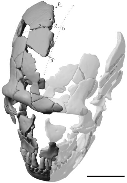
The St. Césaire 1 Neanderthal skeleton of a young adult individual is unique in its association with Châtelperronian artifacts from a level dated to ca. 36,000 years ago. Computer-tomographic imaging and computer-assisted reconstruction of the skull revealed a healed fracture in the cranial vault. When paleopathological and forensic diagnostic standards are applied, the bony scar bears direct evidence for the impact of a sharp implement, which was presumably directed toward the individual during an act of interpersonal violence. These findings add to the evidence that Neanderthals used implements not only for hunting and food processing, but also in other behavioral contexts. It is hypothesized that the high intra-group damage potential inherent to weapons might have represented a major factor during the evolution of hominid social behavior.


Figures

Figure 1
Figure 1
Computerized reconstruction of the St. Césaire 1 Neanderthal skull (mirror-imaged completed parts are transparent) showing the bony scar in the right apical cranial vault. The skull is seen from the direction in which the hypothetical blow was exerted. Arrows indicate the anteroposterior (a and p) extent of the preserved lateral border of the injury (c, coronal suture; b, bregma; *, location of the coronal cross section through the injury (see Fig. 2b); ×, location of the parasagittal cross section through the coronal suture (see Fig. 3c); the dotted line indicates the reconstructed position of the midsagittal plane of the cranial vault). Note oblique, off-midline position of the scar. (Scale bar = 5 cm.)
Figure 2
Figure 2
Anatomy of a blow. (a) Mediolateral view of the right border of the St. Césaire 1 cranial vault injury (same symbols as in Fig. 1: a and p, anterior and posterior delimitations of the scar, c, position of the coronal suture, and *, location of the cross section in b. The drawing indicates areas of bone remodeling (gray) and areas affected by postmortem fracturing (hatched). (b and c) Comparative morphology of the injured vault bones in St. Césaire 1 (b) and a specimen from a medieval graveyard (13) exhibiting a healed scar resulting from sharp trauma on the left frontal bone (c) (both specimens in anteroposterior view, coronal cross sections); the CT sections show bone remodeling at the margin of the injured external lamina (*) and a space (▵) between the dislocated internal lamina and the diploë, probably filled by connective tissue. (Scale bar = 5 cm.)
Figure 3
Figure 3
Comparative cross-sectional morphology of bone injuries and sutures. (a and b) Healed scars of St. Césaire 1 and a medieval specimen (same coronal sections as in Fig. 2 b and c). (c) St. Césaire 1 coronal suture (parasagittal section through × in Fig. 1). (d) Metopic suture in a recent specimen (coronal section). (e) Large occipital trepanation during early phases of healing [Ensisheim skull (14), coronal section]. (f) Healed occipital trepanation in a recent specimen (coronal section). Note apposition of dense cortical bone at the injured borders, resulting in a smooth contour, as opposed to the less dense and corrugated aspect of the sutural borders. (Scale bar = 5 cm.)

Similar articles

Cited by

References

    1. Lévêque F, Vandermeersch B. C R Acad Sci. 1980;291:187–189.
    1. Valladas H, Geneste J-M, Joron J-L, Chadelle J-P. Nature (London) 1986;322:335–344. - PubMed
    1. Mercier N, Valladas H, Joron J L, Reyss J L, Lévêque F, Vandermeersch B. Nature (London) 1991;351:737–739. - PubMed
    1. Hublin J J, Spoor F, Braun M, Zonneveld F, Condemi S. Nature (London) 1996;381:224–226. - PubMed
    1. Vandermeersch B. Bull Mém Soc Anthropol Paris. 1984;1:191–196.

Publication types

LinkOut - more resources