

From intestine to muscle: nuclear reprogramming through defective cloned embryos
- PMID: 11972029
- PMCID: PMC122901
- DOI: 10.1073/pnas.082112099
From intestine to muscle: nuclear reprogramming through defective cloned embryos
Abstract
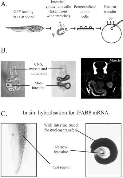
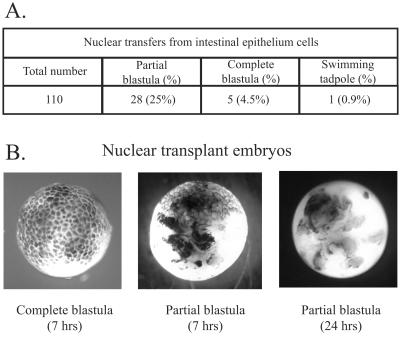
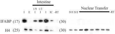
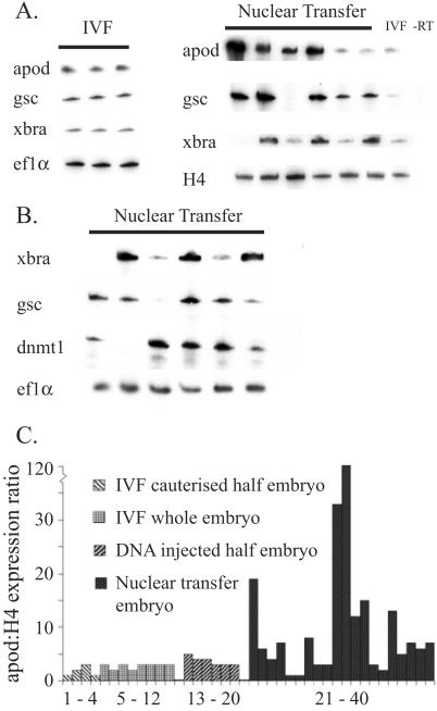
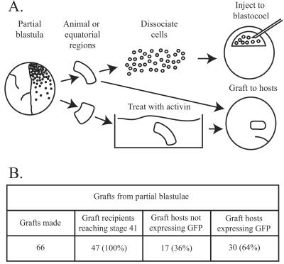
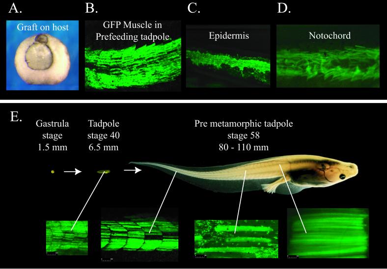
Nuclear transplantation is one of the very few ways by which the genetic content and capacity for nuclear reprogramming can be assessed in individual cells of differentiated somatic tissues. No more than 6% of the cells of differentiated tissues have thus far been shown to have nuclei that can be reprogrammed to elicit the formation of unrelated cell types. In Amphibia, about 25% of such nuclear transfers form morphologically abnormal partial blastulae that die within 24 h. We have investigated the genetic content and capacity for reprogramming of those nuclei that generate partial blastulae, using as donors the intestinal epithelium cells of feeding Xenopus larvae. We have analyzed single nuclear transplant embryos obtained directly from intestinal tissue, thereby avoiding any genetic or epigenetic changes that might accumulate during cell culture. The expression of the intestine-specific gene intestinal fatty acid binding protein is extinguished by at least 10(4) times, within a few hours of nuclear transplantation. At the same time several genes that are normally expressed only in early embryos are very strongly activated in nuclear transplant embryos, but to an unregulated extent. Remarkably, cells from intestine-derived partial blastulae, when grafted to normal host embryos, contribute to several host tissues and participate in the normal 100-fold increase in axial muscle over several months. Thus, cells of defective cloned embryos unable to survive for more than 1 day can be reprogrammed to participate in new directions of differentiation and to maintain indefinite growth, despite the abnormal expression of early genes.
Figures
Similar articles
-
Nuclear reprogramming and stem cell creation.Proc Natl Acad Sci U S A. 2003 Sep 30;100 Suppl 1(Suppl 1):11819-22. doi: 10.1073/pnas.1834207100. Epub 2003 Aug 14. Proc Natl Acad Sci U S A. 2003. PMID: 12920185 Free PMC article. Review.
-
Nuclear transplantation in Xenopus.Methods Mol Biol. 2006;325:1-9. doi: 10.1385/1-59745-005-7:1. Methods Mol Biol. 2006. PMID: 16761714
-
Nuclear cloning, epigenetic reprogramming and cellular differentiation.Novartis Found Symp. 2005;265:107-18; discussion 118-28. Novartis Found Symp. 2005. PMID: 16050253 Review.
-
Transcription of muscle-specific actin genes in early Xenopus development: nuclear transplantation and cell dissociation.Cell. 1984 Oct;38(3):691-700. doi: 10.1016/0092-8674(84)90264-2. Cell. 1984. PMID: 6488316
-
From nuclear transfer to nuclear reprogramming: the reversal of cell differentiation.Annu Rev Cell Dev Biol. 2006;22:1-22. doi: 10.1146/annurev.cellbio.22.090805.140144. Annu Rev Cell Dev Biol. 2006. PMID: 16704337 Review.
Cited by
-
A method for generating transgenic frog embryos.Methods Mol Biol. 2008;461:447-66. doi: 10.1007/978-1-60327-483-8_31. Methods Mol Biol. 2008. PMID: 19030816 Free PMC article. Review. No abstract available.
-
From embryonic stem cells to iPS - an ethical perspective.Cell Prolif. 2011 Apr;44 Suppl 1(Suppl 1):70-84. doi: 10.1111/j.1365-2184.2010.00723.x. Cell Prolif. 2011. PMID: 21481047 Free PMC article. No abstract available.
-
Embryonic death and the creation of human embryonic stem cells.J Clin Invest. 2004 Nov;114(9):1184-6. doi: 10.1172/JCI23065. J Clin Invest. 2004. PMID: 15520846 Free PMC article. Review.
-
Abnormal gene expression in cloned mice derived from embryonic stem cell and cumulus cell nuclei.Proc Natl Acad Sci U S A. 2002 Oct 1;99(20):12889-94. doi: 10.1073/pnas.192433399. Epub 2002 Sep 16. Proc Natl Acad Sci U S A. 2002. PMID: 12235366 Free PMC article.
-
The first half-century of nuclear transplantation.Proc Natl Acad Sci U S A. 2003 Jul 8;100(14):8048-52. doi: 10.1073/pnas.1337135100. Epub 2003 Jun 23. Proc Natl Acad Sci U S A. 2003. PMID: 12821779 Free PMC article.
References
Publication types
MeSH terms
LinkOut - more resources
Full Text Sources
Other Literature Sources
Medical
