. 2002 Apr 30;99(9):6240-5.
doi: 10.1073/pnas.082515999. Epub 2002 Apr 23.

Otoancorin, an inner ear protein restricted to the interface between the apical surface of sensory epithelia and their overlying acellular gels, is defective in autosomal recessive deafness DFNB22

Affiliations

Otoancorin, an inner ear protein restricted to the interface between the apical surface of sensory epithelia and their overlying acellular gels, is defective in autosomal recessive deafness DFNB22

Ingrid Zwaenepoel et al. Proc Natl Acad Sci U S A. .

Abstract

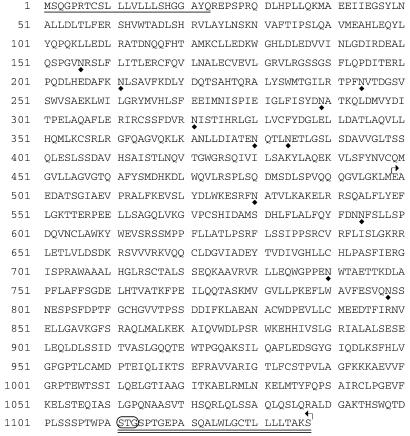
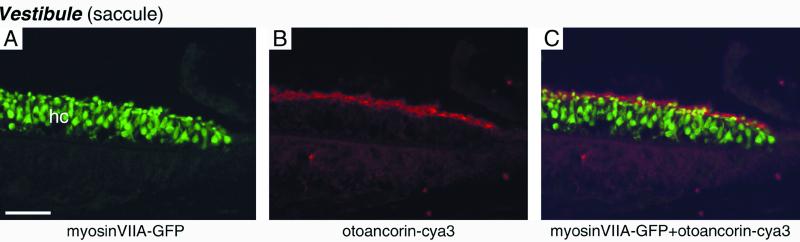
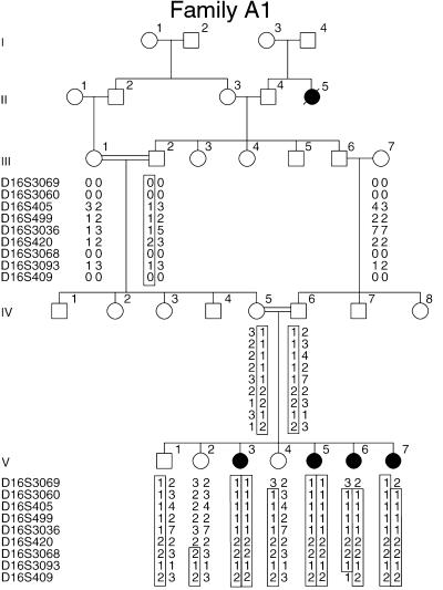
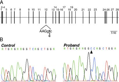
A 3,673-bp murine cDNA predicted to encode a glycosylphosphatidylinositol-anchored protein of 1,088 amino acids was isolated during a study aimed at identifying transcripts specifically expressed in the inner ear. This inner ear-specific protein, otoancorin, shares weak homology with megakaryocyte potentiating factor/mesothelin precursor. Otoancorin is located at the interface between the apical surface of the inner ear sensory epithelia and their overlying acellular gels. In the cochlea, otoancorin is detected at two attachment zones of the tectorial membrane, a permanent one along the top of the spiral limbus and a transient one on the surface of the developing greater epithelial ridge. In the vestibule, otoancorin is present on the apical surface of nonsensory cells, where they contact the otoconial membranes and cupulae. The identification of the mutation (IVS12+2T>C) in the corresponding gene OTOA in one consanguineous Palestinian family affected by nonsyndromic recessive deafness DFNB22 assigns an essential function to otoancorin. We propose that otoancorin ensures the attachment of the inner ear acellular gels to the apical surface of the underlying nonsensory cells.


Figures

Figure 1
Figure 1
RT-PCR analysis of Otoa expression in mouse tissues. (A) Amplification products obtained with B4D7 primers located in the 5′UTR (B4d7ProF) and the last exon (B4d7ProR). A 3,336-bp product is observed only in the inner ear. (B) Products obtained with Hprt primers (positive control). ± indicates presence (+) or absence (−) of reverse transcriptase in the cDNA synthesis reaction. M, 1-kb DNA ladder.
Figure 2
Figure 2
Amino acid sequence of mouse otoancorin. The putative signal peptide is underlined. The characteristic C-terminal signal sequence for addition of a GPI anchor is doubly underlined, with the predicted cleavage site circled. Potential N-glycosylation sites are indicated below the sequence by a diamond-shaped symbol. The region of homology with MPF/mesothelin precursors is delineated by broken arrows.
Figure 3
Figure 3
Distribution of otoancorin in the mouse cochlea revealed by immunofluorescence microscopy. (A) A profile of the cochlear duct at embryonic day 16.5. (B) High magnification of a cross section of the spiral limbus (SL) at P3, showing detail of interdental cells (ic). (C) Section of the organ of Corti at P6. (Inset) A merged image showing a detailed view of the otoancorin-cya3 staining (red) relative to GFP-labeled hair cells (green) in an M3H11-R mouse at P6. GER, greater epithelial ridge; ic, interdental cells; ihc, inner hair cell; IS, inner sulcus; LER, lesser epithelial ridge; ohc, outer hair cells; RM, Reissner's membrane; SL, spiral limbus; TM, tectorial membrane. (Bar = A, B, 30 μm; C, 60 μm.)
Figure 4
Figure 4
Distribution of otoancorin in the saccule of a M3H11-R mouse at P8 revealed by immunofluorescence microscopy. (A–C) Section of the saccular macula. (A) Myosin VIIA-GFP labeling. The sensory hair cells (hc) are labeled in green. (B) Otoancorin-cya3 staining (red). (C) Merged images from A and B. (Bar = 45 μm.)
Figure 5
Figure 5
Distribution of otoancorin in the cochlea of α-tectorin mutant mice (TectaΔENT/ΔENT mice). Immunohistofluorescence at P8 (A) and adult stage (B). Immunoelectron microscopy at P7, showing the ultrastuctural distribution of otoancorin at the apical surface of the spiral limbus (C) and at the apical surface of border epithelial cells (arrowhead in A) lying medial to IHCs (D). Some gold particles are indicated by arrows. (Bars = A and B, 50 μm; C and D, 200 nm.)
Figure 6
Figure 6
Genetic linkage analysis of the DFNB22-affected consanguineous family A1. Segregation analysis with polymorphic microsatellite markers located on chromosome 16 limited the DFNB22 locus interval to between D16S3069 and D16S409. Individuals with prelingual deafness are indicated by filled symbols and unaffected individuals by open symbols. “0” indicates ambiguous positioning of the allele on the gel. The haplotype associated with the mutated DFNB22 allele is boxed.
Figure 7
Figure 7
Detection of the IVS12+2T>C mutation in family A1. (A) Schematic representation of the exon/intron structure of OTOA. Coding exons are indicated by vertical lines. The IVS12+2T>C mutation identified in the affected DFNB22 family A1 is indicated below. Exonic sequence is represented in uppercase letters, intronic sequence by lowercase letters. (B) Sequence analysis of the coding strand at the exon 12/intron 12 junction in a normal control and an affected child. The T to C transition in the affected child is indicated (arrowhead).

References

    1. Zheng J, Shen W, He D Z, Long K B, Madison L D, Dallos P. Nature (London) 2000;405:149–155. - PubMed
    1. Legan P K, Rau A, Keen J N, Richardson G P. J Biol Chem. 1997;272:8791–8801. - PubMed
    1. Cohen-Salmon M, El-Amraoui A, Leibovici M, Petit C. Proc Natl Acad Sci USA. 1997;94:14450–14455. - PMC - PubMed
    1. Verpy E, Leibovici M, Petit C. Proc Natl Acad Sci USA. 1999;96:529–534. - PMC - PubMed
    1. Wang Y, Kowalski P E, Thalmann I, Ornitz D M, Mager D L, Thalmann R. Proc Natl Acad Sci USA. 1998;95:15345–15450. - PMC - PubMed

Publication types

MeSH terms

Associated data