. 2002 Apr 30;99(9):6316-21.
doi: 10.1073/pnas.092128499. Epub 2002 Apr 23.

YgbQ, a cell division protein in Escherichia coli and Vibrio cholerae, localizes in codependent fashion with FtsL to the division site

Affiliations

YgbQ, a cell division protein in Escherichia coli and Vibrio cholerae, localizes in codependent fashion with FtsL to the division site

Nienke Buddelmeijer et al. Proc Natl Acad Sci U S A. .

Abstract

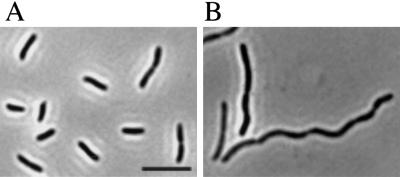
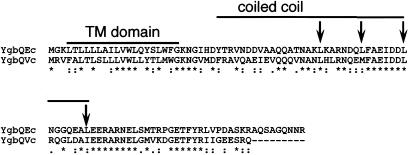
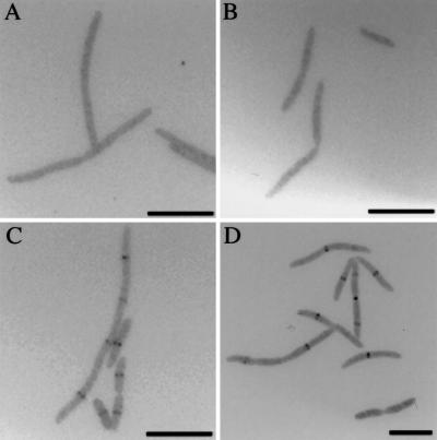
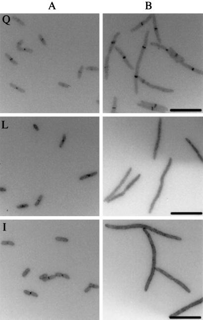
YgbQ is a cell division protein in Escherichia coli and Vibrio cholerae. In E. coli the ygbQ gene was discovered as a result of a computer search of the E. coli genome designed to find potential interacting partners for cell division protein FtsL. In V. cholerae, ygbQ was identified as an essential gene by using a transposon that fuses genes to an arabinose promoter. The role of YgbQ in cell division is supported by the following. Cells depleted of YgbQ in both organisms form long filaments, but DNA segregation is not affected. YgbQ localizes to the constriction site in wild-type E. coli cells. Localization of E. coli YgbQ to the constriction site depends on cell division proteins FtsQ and FtsL but not FtsW and FtsI, placing YgbQ in the sequential dependency order of proteins localizing to the division site. Localization of green fluorescent protein-FtsL also depends on YgbQ, indicating that FtsL and YgbQ colocalize to the division site in E. coli. Our results show colocalization of proteins to the bacterial midcell in E. coli and raise the possibility that these proteins interact in a coiled-coil structure.


Figures

Figure 1
Figure 1
Phenotype of ygbQ depletion strain of V. cholerae. (A) Image of cells grown with arabinose to express ygbQ. (B) Image of cells grown with glucose to deplete YgbQ. (Scale bar, 10 μm.)
Figure 2
Figure 2
CLUSTALW 1.81 alignment of YgbQ of E. coli (Ec) and V. cholerae (Vc). The leucine residues that form the leucine zipper-like motif in the periplasmic domain of YgbQ of E. coli are indicated with arrows. The overlined regions represent the putative transmembrane (TM) domain (residues 4–22) and the coiled-coil domain (residues 29–67) of YgbQ. Conserved amino acid residues are indicated by asterisks.
Figure 3
Figure 3
Localization of GFP fusions to YgbQ in FtsQ (A), FtsL (B), FtsW (C), and FtsI (D) depletion strains. (Scale bars, 10 μm.)
Figure 4
Figure 4
Localization of GFP fusions to FtsQ (Q), FtsL (L), and FtsI (I) in a ygbQec depletion strain. (A) Localization of GFP-fusion proteins in cells grown with arabinose to express ygbQ. (B) Localization of GFP-fusion proteins in cells grown with glucose to deplete ygbQ. (Scale bars, 10 μm.)
Figure 5
Figure 5
Detection of wild-type levels FtsL in membranes of a ygbQ null strain (NB946). Lanes 1 and 2, NB946 grown in arabinose to express ygbQ for 3 and 4 h, respectively; lanes 3 and 4, NB946 grown in glucose to deplete ygbQ for 3 and 4 h, respectively.

References

    1. Rothfield L, Justice S, Garcia-Lara J. Annu Rev Genet. 1999;33:423–448. - PubMed
    1. Margolin W. FEMS Microbiol Rev. 2000;24:531–548. - PubMed
    1. Chen J C, Beckwith J. Mol Microbiol. 2001;42:395–413. - PubMed
    1. Ghigo J-M, Beckwith J. J Bacteriol. 2000;182:116–129. - PMC - PubMed
    1. Guzman L-M, Barondess J, Beckwith J. J Bacteriol. 1992;174:7716–7728. - PMC - PubMed

Publication types

MeSH terms

Substances

LinkOut - more resources