. 2002 Apr 30;99(9):6181-6.
doi: 10.1073/pnas.092141999. Epub 2002 Apr 23.

MIP-1alpha, MIP-1beta, RANTES, and ATAC/lymphotactin function together with IFN-gamma as type 1 cytokines

Affiliations

MIP-1alpha, MIP-1beta, RANTES, and ATAC/lymphotactin function together with IFN-gamma as type 1 cytokines

Brigitte G Dorner et al. Proc Natl Acad Sci U S A. .

Abstract

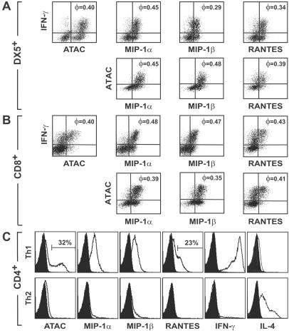
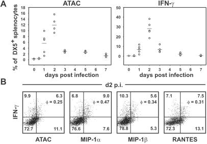
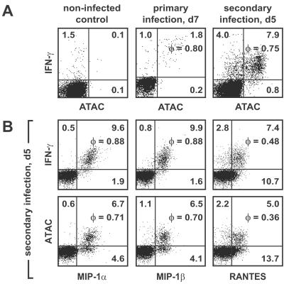
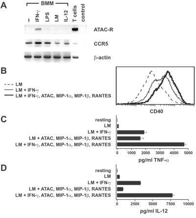
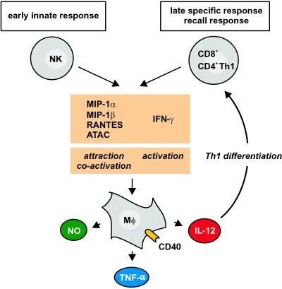
We analyzed for the first time the expression of chemokines in subpopulations of the murine immune system at the single-cell level. We demonstrate in vitro and in a model of murine listeriosis that macrophage inflammatory protein (MIP)-1alpha, MIP-1beta, regulated on activation normal T cell expressed and secreted (RANTES), and activation-induced, T cell-derived, and chemokine-related cytokine (ATAC)/lymphotactin are cosecreted to a high degree with IFN-gamma by activated individual natural killer (NK), CD8(+) T, and CD4(+) T helper 1 (Th1) cells. Functionally, ATAC and the CC chemokines cooperate with IFN-gamma in the up-regulation of CD40, IL-12, and tumor necrosis factor-alpha, molecules playing a central role in the effector phase of macrophages. Our data indicate that (i) MIP-1alpha, MIP-1beta, RANTES, and ATAC are not only chemoattractants but also coactivators of macrophages, (ii) MIP-1alpha, MIP-1beta, RANTES, and ATAC constitute together with IFN-gamma a group of "type 1 cytokines," and (iii) these cytokines act together as a functional unit that is used by NK cells in the innate phase and then "handed over" to CD8(+) T cells in the antigen-specific phase of the immune defense, thus bridging the two components of a Th1 immune reaction.


Figures

Figure 1
Figure 1
ATAC, MIP-1α, MIP-1β, RANTES, and IFN-γ are cosecreted in polyclonally activated NK cells, CD8+ T cells, and Th1-differentiated CD4+ T cells. C57BL/6 splenocytes were stimulated with phorbol 12-myristate 13-acetate, ionomycin, and brefeldin A for 6 h, stained for DX5 or CD8, fixed, and counterstained with the respective cytokine/chemokine reagents. (A) Gate on DX5+ cells. (B) Gate on CD8+ cells. The correlation coefficient φ is indicated in the right upper quadrants. Shown is representative of four experiments. (C) OVA-specific Th1 or Th2 cells were restimulated after 2 weeks of differentiation with phorbol 12-myristate 13-acetate, ionomycin, and brefeldin A for 6 h and analyzed for the expression of ATAC, the CC chemokines, IFN-γ, and IL-4. Black profiles indicate isotype controls. The data given were obtained in one representative experiment out of two.
Figure 2
Figure 2
Correlated secretion of IFN-γ with ATAC, MIP-1α, MIP-1β, and RANTES by NK cells in the innate phase of L. monocytogenes infection. (A) Time course of ATAC and IFN-γ secretion in splenic DX5+ cells. BALB/c mice were infected with L. monocytogenes (five mice per group), and the spleens were removed at various time points. Splenocytes were incubated with brefeldin A for 5 h without restimulation and analyzed for the expression of ATAC and IFN-γ by flow cytometry. Shown is the percentage of DX5+ cells secreting ATAC or IFN-γ in the course of listeriosis. (B) On day 2 (d2) p.i. splenocytes from individual Listeria-infected BALB/c mice were stained for DX5 and counterstained for IFN-γ versus ATAC, MIP-1α, MIP-1β, or RANTES. The percentage of cells in each quadrant and the correlation coefficient φ for each staining pair are indicated. Analysis of one representative animal out of eight is shown.
Figure 3
Figure 3
Cosecretion of ATAC, MIP-1α, MIP-1β, RANTES, and IFN-γ by antigen-specific CD8+ T cells during the adaptive phase of L. monocytogenes infection. BALB/c mice were infected with a sublethal dose of L. monocytogenes. On day 7 (d7) p.i. of primary infection or day 5 (d5) p.i. of a secondary infection, splenocytes of individual mice were restimulated with the Listeria-specific peptide LLO91–99 for 6 h in the presence of brefeldin A. Cells were stained for CD8, fixed, and counterstained for IFN-γ, ATAC, MIP-1α, MIP-1β, or RANTES. (A) Cosecretion of IFN-γ with ATAC by Listeria-specific CD8+ T cells in primary and secondary infection. (B) Cosecretion of IFN-γ and ATAC with MIP-1α, MIP-1β, and RANTES by Listeria-specific CD8+ T cells in secondary infection. Analysis of one representative animal out of six is shown.
Figure 4
Figure 4
Functional effects of the type 1 chemokines and IFN-γ on macrophages. (A) BMMs were exposed to IFN-γ, lipopolysaccharide (LPS), or IL-12 or were infected with live L. monocytogenes as described in Materials and Methods. After 24 h of culture, total RNA was analyzed by RT-PCR for the ATAC receptor, CCR5, and β-actin. RNA from purified (99%) CD8+ T cells (T cells) was used as a positive control, and samples without mRNA template were used as a negative control (control). (B) BMMs were infected with Listeria and were either left untreated (LM, dashed line) or treated additionally with IFN-γ alone (thin line) or a combination of IFN-γ and ATAC, MIP-1α, MIP-1β, and RANTES (thick line). After 24 h, BMMs were collected and analyzed for CD40 cell surface expression by using flow cytometry. One representative experiment out of five is shown. (C and D) BMMs were either left untreated (resting) or infected with L. monocytogenes for 1 h. Infected BMMs were either cultured in medium or stimulated with cytokines as indicated. After 24 h, supernatants were harvested and tested for the presence of TNF-α (C) and IL-12 (D). The values represent means of duplicates (TNF-α and IL-12) ± SD. One representative out of four experiments is shown.
Figure 5
Figure 5
Model for the role of the type 1 chemokines and IFN-γ as a functional unit in the course of a Th1-inducing infection. MIP-1α, MIP-1β, RANTES, ATAC, and IFN-γ are cosecreted early by NK cells and later by CD8+ T/CD4+ Th1 cells. In both phases, the type 1 chemokines attract and, together with IFN-γ, coactivate macrophages to release NO, TNF-α, and IL-12. The chemokine group MIP-1α, MIP-1β, RANTES, and ATAC thus constitute together with the cytokine IFN-γ a functional unit that is used both by cells of the innate and adaptive immunity to drive the type 1 immune reaction in vivo.

References

    1. Zlotnik A, Yoshie O. Immunity. 2000;12:121–127. - PubMed
    1. Moser B, Loetscher P. Nat Immunol. 2001;2:123–128. - PubMed
    1. Baggiolini M. Adv Immunol. 1994;55:97–179. - PubMed
    1. Murphy P M, Baggiolini M, Charo I F, Hebert C A, Horuk R, Matsushima K, Miller L H, Oppenheim J J, Power C A. Pharmacol Rev. 2000;52:145–176. - PubMed
    1. Uguccioni M, D'Apuzzo M, Loetscher M, Dewald B, Baggiolini M. Eur J Immunol. 1995;25:64–68. - PubMed

Publication types

MeSH terms