. 2002 Apr 30;99(9):6234-9.
doi: 10.1073/pnas.092637699. Epub 2002 Apr 23.

Dilated cardiomyopathy in mice deficient for the lysosomal cysteine peptidase cathepsin L

Affiliations

Dilated cardiomyopathy in mice deficient for the lysosomal cysteine peptidase cathepsin L

Jörg Stypmann et al. Proc Natl Acad Sci U S A. .

Abstract

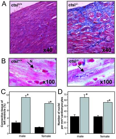
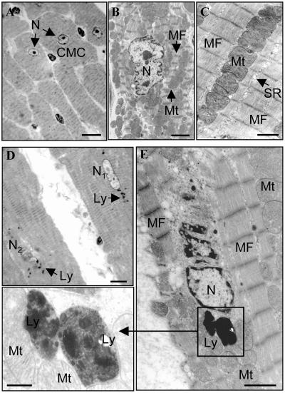
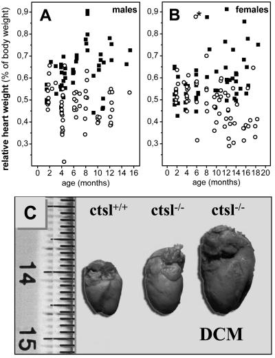
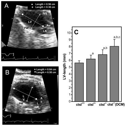
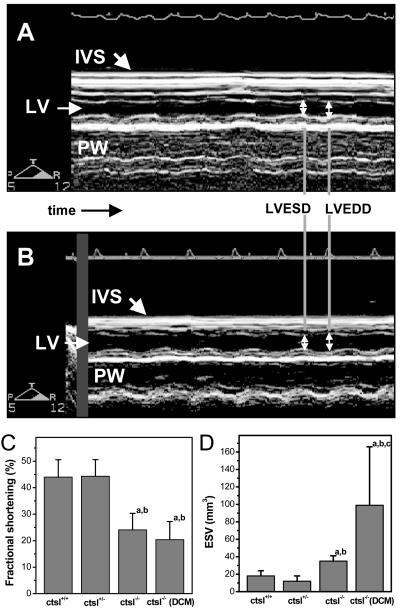
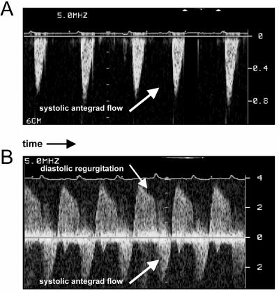
Dilated cardiomyopathy is a frequent cause of heart failure and is associated with high mortality. Progressive remodeling of the myocardium leads to increased dimensions of heart chambers. The role of intracellular proteolysis in the progressive remodeling that underlies dilated cardiomyopathy has not received much attention yet. Here, we report that the lysosomal cysteine peptidase cathepsin L (CTSL) is critical for cardiac morphology and function. One-year-old CTSL-deficient mice show significant ventricular and atrial enlargement that is associated with a comparatively small increase in relative heart weight. Interstitial fibrosis and pleomorphic nuclei were found in the myocardium of the knockout mice. By electron microscopy, CTSL-deficient cardiomyocytes contained multiple large and apparently fused lysosomes characterized by storage of electron-dense heterogeneous material. Accordingly, the assessment of left ventricular function by echocardiography revealed severely impaired myocardial contraction in the CTSL-deficient mice. In addition, echocardiographic and electrocardiographic findings to some degree point to left ventricular hypertrophy that most likely represents an adaptive response to cardiac impairment. The histomorphological and functional alterations of CTSL-deficient hearts result in valve insufficiencies. Furthermore, abnormal heart rhythms, like supraventricular tachycardia, ventricular extrasystoles, and first-degree atrioventricular block, were detected in the CTSL-deficient mice.


Figures

Figure 1
Figure 1
Histological and histomorphometric analysis of myocardium. (A) Trichrome stain of myocardium of wild-type mice (ctsl+/+) and CTSL-deficient mice (ctsl−/−) at 12 months of age. Connective tissue appears in blue. (B) Large pleomorphic nucleus (P) in CTSL-deficient myocardium as compared with a normal nucleus (N). Masson's trichrome stain at 100-fold resolution. Note the distinct interstitial collagen staining in the absence of CTSL. (C) Histomorphometric determination of left ventricular connective tissue of wild-type (black) and CTSL-deficient (gray) mice. (D) Numeric density of nuclei in cardiomyocytes of wild-type (black) and CTSL-deficient (gray) ventricles. (a) P < 0.001 for comparison of wild-type (10 females, seven males) and knockout (nine females, five males) mice.
Figure 2
Figure 2
Electron microscopic analysis of myocardium in 12-month-old ctsl+/+ and ctsl−/− mice. (A) Low-power view of cardiac muscle cells (CMC) of ctsl+/+ heart showing several typical cylindrical, striated, muscle cells (transversely cut) with a centrally located clear nucleus (N). (B) Medium-power view of single cardiac muscle cell with central clear nucleus (N) surrounded by numerous mitochondria (Mt) and myofibrils (MF). (C) High-power view of cardiac muscle cell cytoplasm showing organized arrays of myofibrils (MF) and closely packed mitochondria (MT) and sarcoplasmic reticulum. Note that lysosomes are not readily apparent in ctsl+/+ tissue. (D) Low-power view of cardiac muscle cells of ctsl−/− heart showing two cylindrical striated muscle cells (longitudinally cut). Several lysosomes (Ly) are distributed around nuclei that may be morphologically normal (N1) or undergoing lysis (N2). (E) Higher-power view of a single cardiac muscle cell in ctsl−/− heart. Large lysosomes (Ly) are distributed close to a dystrophic nucleus (N) and surrounded by numerous mitochondria (Mt) and myofibrils (MF). (Inset) High-power view of lysosomes (Ly) surrounded by myofibrils (MF) and mitochondria (Mt). Lysosomes contain heterogeneous electron-dense materials. (A and D) High-resolution light microscopy, toluidine blue stain. [Scale bars: A and D, 20 μm; B, C, and E (transmission electron microscopy, uranyl acetate, and lead citrate), B, 2 μm; C, 1 μm; E, 1 μm; Inset, 0.3 μm.]
Figure 3
Figure 3
Relative heart weights of CTSL-deficient mice and heart morphology. Relative heart weights of male (A) ctsl−/− (■, n = 52) and ctsl+/+ (○, n = 62) and female (B) ctsl−/− (■, n = 58) and ctsl+/+ (○, n = 69) mice. A 6-month-old wild-type mouse with severely enlarged heart is indicated by *. This mouse presented the only “spontaneous” heart disease seen in the 131 investigated wild-type mice. (C) Hearts of wild-type (ctsl+/+) and CTSL-deficient (ctsl−/−) mice at 12 months of age are shown. About 75% of ctsl−/− hearts show slight to moderate enlargement. The remaining 25% of ctsl−/− hearts exhibit severe enlargement with severe functional impairment, manifest DCM.
Figure 4
Figure 4
In vivo assessment of left ventricular dimensions by echocardiography. (A) Two-dimensional echocardiographic picture of a normal-sized left ventricle in a 12-month-old wild-type mouse. Ao, aortic valve; LV, left ventricle; LA, left atrium; IVS, interventricular septum; PW, posterior wall. (B) Enlarged left ventricle of a 12-month-old CTSL-deficient mouse. (C) Genotype-dependent increase in the length of left ventricle. ctsl+/+, wild-type mice; ctsl+/−, heterozygous mice; ctsl−/−, CTSL-deficient mice; ctsl−/− DCM, CTSL-deficient mice with manifest DCM. (a) P < 0.05 compared with ctsl+/+, (b) P < 0.05 compared with ctsl+/−, (c) P < 0.05 compared with ctsl−/−.
Figure 5
Figure 5
Determination of heart contraction. (A) M-mode echocardiography showing normal contraction of the interventricular septum and the posteriolateral wall in the heart of a wild-type mouse. LV, left ventricle; IVS, interventricular septum; PW, posterior wall; LVEDD, left ventricular end-diastolic diameter; LVESD, left ventricular end-systolic diameter. (B) Reduced contraction of interventricular septum and posteriolateral wall in a CTSL-deficient mouse. (C) Fractional shortening as a measure of heart contraction and (D) left ventricular end-systolic volume in wild-type mice (ctsl+/+), heterozygous mice (ctsl+/−), CTSL-deficient mice (ctsl−/−), and CTSL-deficient mice with manifest DCM (ctsl−/− DCM). (a) P < 0.05 compared with ctsl+/+, (b) P < 0.05 compared with ctsl+/−, (c) P < 0.05 compared with ctsl−/−.
Figure 6
Figure 6
Functional assessment of heart valves by pulse-waved Doppler-echocardiography. (A) Wild-type mouse showing normal antegrade flow through the aortic valve. (B) Aortic regurgitation and high velocity of aortic antegrade flow as hallmarks of aortic valve insufficiency in a ctsl−/− mouse.

Similar articles

Cited by

References

    1. Kamisago M, Sharma S D, DePalma S R, Solomon S, Sharma P, McDonough B, Smoot L, Mullen M P, Woolf P K, Wigle E D, et al. N Engl J Med. 2000;343:1688–1696. - PubMed
    1. Olson T M, Michels V V, Thibodeau S N, Tai Y S, Keating M T. Science. 1998;280:750–752. - PubMed
    1. Li D, Tapscoft T, Gonzalez O, Burch P E, Quinones M A, Zoghbi W A, Hill R, Bachinski L L, Mann D L, Roberts R. Circulation. 1999;100:461–464. - PubMed
    1. Towbin J A, Hejtmancik J F, Brink P, Gelb B, Zhu X M, Chamberlain J S, McCabe E R, Swift M. Circulation. 1993;87:1854–1865. - PubMed
    1. Tsubata S, Bowles K R, Vatta M, Zintz C, Titus J, Muhonen L, Bowles N E, Towbin J A. J Clin Invest. 2000;106:655–662. - PMC - PubMed

Publication types