. 2002 May 1;30(9):1879-85.
doi: 10.1093/nar/30.9.1879.

Charge neutralization and DNA bending by the Escherichia coli catabolite activator protein

Affiliations

Charge neutralization and DNA bending by the Escherichia coli catabolite activator protein

Philip R Hardwidge et al. Nucleic Acids Res. .

Abstract

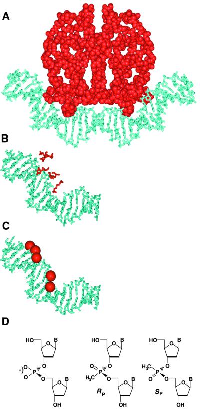
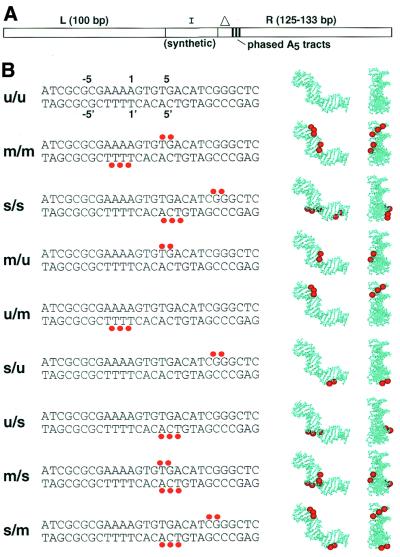
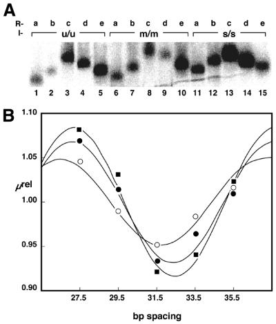
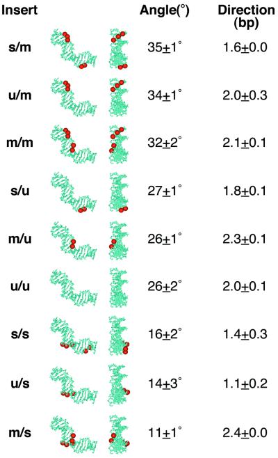
We are interested in the role of asymmetric phosphate neutralization in DNA bending induced by proteins. We describe an experimental estimate of the actual electrostatic contribution of asymmetric phosphate neutralization to the bending of DNA by the Escherichia coli catabolite activator protein (CAP), a prototypical DNA-bending protein. Following assignment of putative electrostatic interactions between CAP and DNA phosphates based on X-ray crystal structures, appropriate phosphates in the CAP half-site DNA were chemically neutralized by methylphosphonate substitution. DNA shape was then evaluated using a semi-synthetic DNA electrophoretic phasing assay. Our results confirm that the unmodified CAP DNA half-site sequence is intrinsically curved by 26 degrees in the direction enhanced in the complex with protein. In the absence of protein, neutralization of five appropriate phosphates increases DNA curvature to 32 degrees (approximately 23% increase), in the predicted direction. Shifting the placement of the neutralized phosphates changes the DNA shape, suggesting that sequence-directed DNA curvature can be modified by the asymmetry of phosphate neutralization. We suggest that asymmetric phosphate neutralization contributes favorably to DNA bending by CAP, but cannot account for the full DNA deformation.


Figures

Figure 1
Figure 1
Asymmetric phosphate neutralization in CAP–DNA complexes. (A) CAP–DNA structure, derived from Schultz et al. (3) and Parkinson et al. (4). CAP is shown in red, DNA in cyan. (B) Left half-site DNA, highlighting the five basic side-chains of CAP (K26, K166, R169, K188 and H199) that contact DNA phosphates [nomenclature from Parkinson et al. (4)]. (C) Modified half-site DNA. Methylphosphonate substitutions shown as red spheres. (D) Structures of anionic phosphodiester linkage (left) and neutral methylphosphonate diastereomeric linkages (right).
Figure 2
Figure 2
Experimental design. (A) Semi-synthetic phasing probes. Phasing probes are trimolecular ligation products, containing a synthetic DNA duplex insert (I) flanked by restriction fragments (L and R). In the present study each strand of the synthetic insert was 24 bases in length. Three phased A5- tracts in the right arms are indicated as vertical black lines. Right arms differ only in the initial distance to the first A5- tract of the array, indicated by the triangle. (B) The DNA duplex inserts (I) under investigation. Red dots indicate sites of methylphosphonate substitution. Top strand sequences are shown 5′→3′ (left to right). Single letter codes (left) refer to substitutions on the top/bottom strands, respectively, where u refers to an unmodified DNA strand, m refers to a DNA strand bearing methylphosphonate residues on phosphates contacted by basic amino acids in the crystal structure and s refers to strands bearing methylphosphonates as in m, but shifted 5 bp to the right. Coordinates above and below u/u DNA strands indicate the numbering of DNA bases in the CAP half-site, as defined by Parkinson et al. (4). Molecular graphics at right depict neutralized phosphates mapped onto the DNA structure extracted from the X-ray model (left of pair is side view, right of pair is same molecule viewed in the plane of the figure, from right).
Figure 3
Figure 3
Phasing assay data. (A) Image obtained after native polyacrylamide gel electrophoresis of phasing probes containing duplexes u/u, m/m and s/s. Identities of right phasing arms are shown, with a, b, c, d and e referring to a spacing of 27.5, 29.5, 31.5, 33.5 and 35.5 bp, respectively, between the center of curvature in the proximal A5 -tract and position 1 of the CAP half-site (Fig. 2B). (B) Quantitative analysis of electrophoretic data for inserts u/u (filled circles), m/m (filled squares) and s/s (open circles). The relative mobility of each of the five phasing probes in a given protein complex (µrel) was plotted as a function of the spacing (in base pairs) between the center of curvature in the proximal A5- tract and position 1 of the CAP half-site (Fig. 2B). Data were fit to eqution 2 as described in Materials and Methods.
Figure 4
Figure 4
Results of charge neutralization experiments. DNA inserts are ranked on the basis of apparent curvature in electrophoretic phasing experiments, from most curved (top) to least curved (bottom). Duplex designations are as in Figure 2B, followed by side and end views of the synthetic inserts with neutralizations (red) depicted on the DNA structural model extracted from the CAP/DNA X-ray structures (for clarity). Apparent bend angles were deduced from analysis of electrophoretic phasing data as in Figure 3 and are described as the average and standard deviation of at least two independent experiments. Bend direction is deduced from the curve fitting of phasing data as described in Materials and Methods. Bend direction is defined here as towards the minor groove in a reference frame centered on the indicated base pair position (as defined by the numbering shown in Fig. 2B). For reference, the primary DNA kink in the left half-site of the crystal structure is toward the major groove at position 6.5. The smaller secondary DNA kink is toward the minor groove between positions –1 and 1.

References

    1. Passner J.M. and Steitz,T.A. (1997) The structure of a CAP-DNA complex having two cAMP molecules bound to each monomer. Proc. Natl Acad. Sci. USA, 94, 2843–2847. - PMC - PubMed
    1. Liu-Johnson H.N., Gartenberg,M.R. and Crothers,D.M. (1986) The DNA binding domain and bending angle of the E. coli CAP protein. Cell, 47, 995–1005. - PubMed
    1. Schultz S.C., Shields,G.C. and Steitz,T.A. (1991) Crystal structure of a CAP-DNA complex: the DNA is bent by 90°. Science, 253, 1001–1007. - PubMed
    1. Parkinson G., Wilson,C., Gunasekera,A., Ebright,Y.W., Ebright,R.E. and Berman,H.M. (1996) Structure of the CAP-DNA complex at 2.5 Å resolution: a complete picture of the protein-DNA interface. J. Mol. Biol., 260, 395–408. - PubMed
    1. Kahn J.D. and Crothers,D.M. (1998) Measurement of the DNA bend angle induced by the catabolite activator protein using Monte Carlo simulation of cyclization kinetics. J. Mol. Biol., 276, 287–309. - PubMed

Publication types