. 2002 May 1;30(9):2018-24.
doi: 10.1093/nar/30.9.2018.

Two-piece tmRNA in cyanobacteria and its structural analysis

Affiliations

Two-piece tmRNA in cyanobacteria and its structural analysis

Cyril Gaudin et al. Nucleic Acids Res. .

Abstract

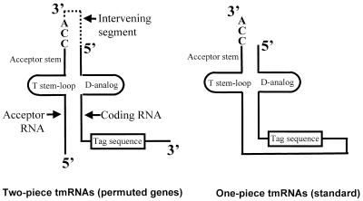
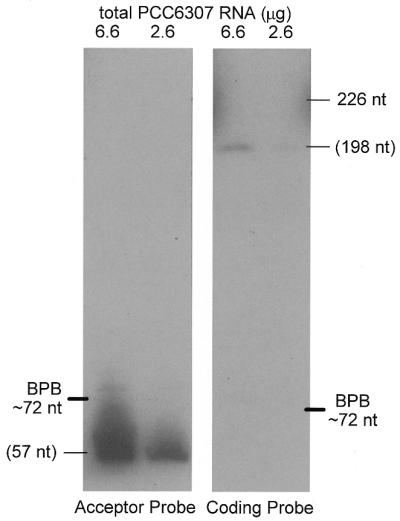
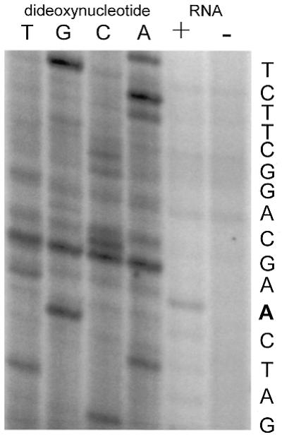
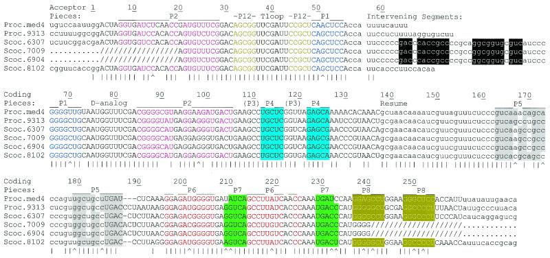
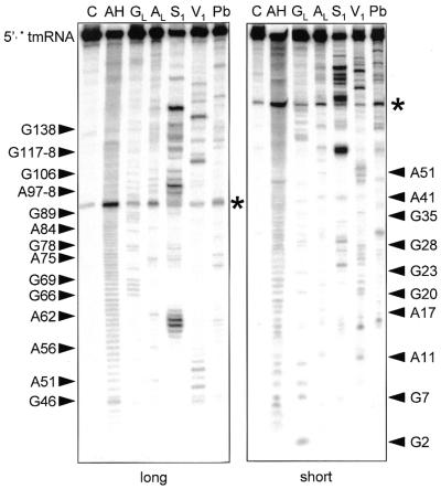
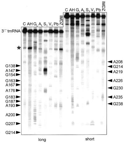
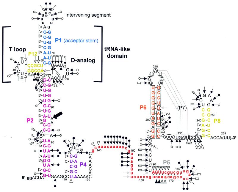
tmRNA acts to rescue stalled bacterial ribosomes while encoding a peptide tag added trans-translationally to the nascent peptide, targeting it for proteolysis. The permuted gene structure found in a group of cyanobacteria is shown to produce a two-piece mature tmRNA, as had been observed previously for the independently permuted gene of alpha-proteobacteria. The pieces have been mapped onto the gene sequence and aligned for the permuted cyanobacterial tmRNA sequences, including four novel sequences. Structural probing and base pair co-variations support a secondary structure model in which two pairings in the tRNA-like domain hold the two pieces together, and the coding piece bearing the tag reading frame additionally contains a single transient pseudoknot and three other stem-loops. This represents a dramatic reduction in pseudoknot number from the five present in one-piece cyanobacterial tmRNA.


Figures

Figure 1
Figure 1
Structural relationship between one-piece and two-piece tmRNAs. In the two-piece tmRNA the structure contains two distinct RNA molecules, the acceptor RNA and the coding RNA (14); the processed portion of the precursor is denoted by dotted lines.
Figure 2
Figure 2
Two tmRNA pieces. Northern blot analysis of PCC6307 RNA using separate probes for the two tmRNA pieces. Sizes in parentheses were estimated by calibration with standards.
Figure 3
Figure 3
Determination of the acceptor piece 5′ end. Primer extension in the presence, but not absence, of total PCC6307 RNA produced a product that maps the 5′ end of tmRNA acceptor piece RNA to the adenosine highlighted in the gene sequence at right.
Figure 4
Figure 4
Alignment of two-piece cyanobacterial tmRNAs. Intervening and 5′ and 3′ flanking segments, tag reading frames and the CCA tails expected to be added after transcription are in lower case. Slashes show segments of unverified sequence where PCR was primed. Vertical lines mark positions conserved among all sequences and carats mark positions where proposed pairings (color coding) exhibit Watson–Crick base pair co-variation. Pairings are numbered according to an earlier proposal (14), but modified to reflect probing data (see below) that revealed extensions of P5 and P6 and that P3 does not form. Pm, P.marinus; Sc, Synechococcus. Numbering is for the pre-tmRNA transcript used for probing experiments.
Figure 5
Figure 5
Structural probing of the 5′ half of a two-piece tmRNA precursor transcript. Autoradiogram of cleavage products of 5′-labeled RNA by imidazole, lead and nucleases S1 and V1. Lanes C, incubation controls; lanes GL, RNase T1 hydrolysis ladder; lanes AL, RNase U2 hydrolysis ladder; lanes AH, alkaline hydrolysis ladder. The sequence is indexed at the sides. Asterisks show a strong spontaneous degradation site.
Figure 6
Figure 6
Structural probing of the 3′-half. Autoradiogram of cleavage products of 3′-labeled RNA with indications as in Figure 5.
Figure 7
Figure 7
Secondary structure model of the P.marinus MED4 pre-tmRNA, based on probing and phylogenetic data, showing the probing results. Triangles are V1 cuts; arrows capped by a circle are S1 cuts; uncapped arrows are lead acetate cuts; arrows capped by a rectangle are imidazole-induced cleavages. Intensities of cuts and cleavages are proportional to the darkness of the symbols: open, stippled and filled for weak, medium and strong cuts, respectively. A consistently observed degradation site (Fig. 5) is indicated by a bold arrow. Circled nucleotides are accessible to DMS or CMCT, squared nucleotides are protected. Structural domains P1, P2, P6, P8 and P12 are color coded as in Figure 4. The 3′-terminal nucleotides in parentheses are not present in the synthetic, but the precise length and 3′-terminal sequence of the coding piece is unknown. Lower case represents 5′ nucleotides added to improve in vitro transcription, the intervening segment and the tag reading frame (in red).

References

    1. Karzai A.W., Roche,E.D. and Sauer,R.T. (2000) The SsrA-SmpB system for protein tagging, directed degradation and ribosome rescue. Nature Struct. Biol., 7, 449–455. - PubMed
    1. Gillet R. and Felden,B. (2001) Emerging views on tmRNA mediated protein tagging and ribosome rescue. Mol. Microbiol., 42, 879–885. - PubMed
    1. Karzai A.W., Susskind,M.M. and Sauer,R.T. (1999) SmpB, a unique RNA-binding protein essential for the peptide-tagging activity of SsrA (tmRNA). EMBO J., 18, 3793–3799. - PMC - PubMed
    1. Rudinger-Thirion J., Giege,R. and Felden,B. (1999) Aminoacylated tmRNA from Escherichia coli interacts with prokaryotic elongation factor Tu. RNA, 5, 1–4. - PMC - PubMed
    1. Wower I.K., Zwieb,C.W., Guven,S.A. and Wower,J. (2000) Binding and cross-linking of tmRNA to ribosomal protein S1, on and off the Escherichia coli ribosome. EMBO J., 19, 6612–6621. - PMC - PubMed

Publication types

Associated data