. 2002 May;184(10):2603-13.
doi: 10.1128/JB.184.10.2603-2613.2002.

Functional characterization and regulation of gadX, a gene encoding an AraC/XylS-like transcriptional activator of the Escherichia coli glutamic acid decarboxylase system

Affiliations

Functional characterization and regulation of gadX, a gene encoding an AraC/XylS-like transcriptional activator of the Escherichia coli glutamic acid decarboxylase system

Angela Tramonti et al. J Bacteriol. 2002 May.

Abstract

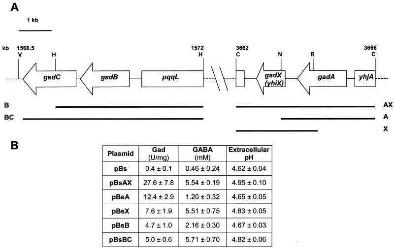
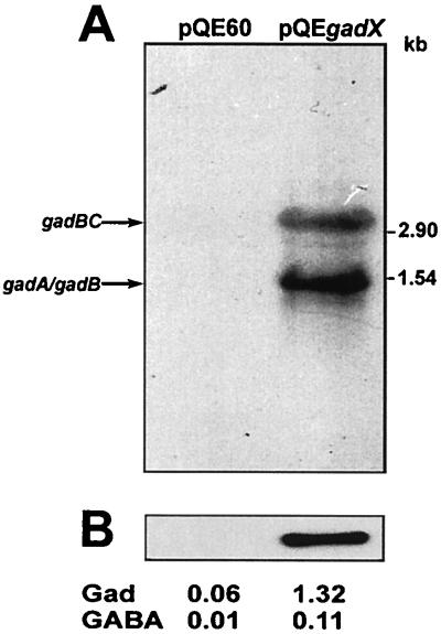
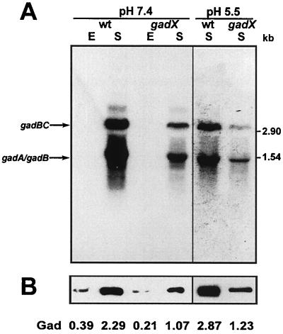
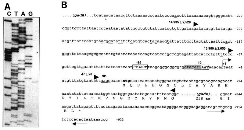
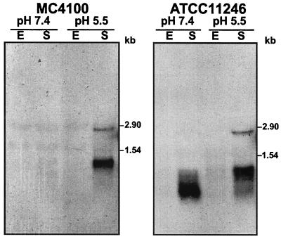
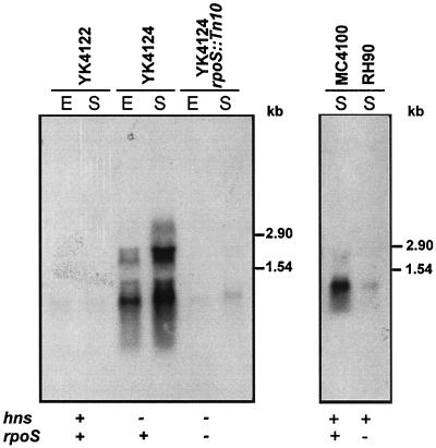
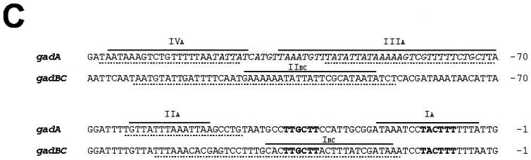
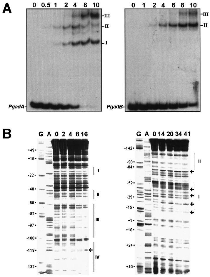
The Escherichia coli chromosome contains two distantly located genes, gadA and gadB, which encode biochemically undistinguishable isoforms of glutamic acid decarboxylase (Gad). The Gad reaction contributes to pH homeostasis by consuming intracellular H(+) and producing gamma-aminobutyric acid. This compound is exported via the protein product of the gadC gene, which is cotranscribed with gadB. Here we demonstrate that transcription of both gadA and gadBC is positively controlled by gadX, a gene downstream of gadA, encoding a transcriptional regulator belonging to the AraC/XylS family. The gadX promoter encompasses the 67-bp region preceding the gadX transcription start site and contains both RpoD and RpoS putative recognition sites. Transcription of gadX occurs in neutral rich medium upon entry into the stationary phase and is increased at acidic pH, paralleling the expression profile of the gad structural genes. However, P(T5)lacO-controlled gadX expression in neutral rich medium results in upregulation of target genes even in exponential phase, i.e., when the gad system is normally repressed. Autoregulation of the whole gad system is inferred by the positive effect of GadX on the gadA promoter and gadAX cotranscription. Transcription of gadX is derepressed in an hns mutant and strongly reduced in both rpoS and hns rpoS mutants, consistent with the expression profile of gad structural genes in these genetic backgrounds. Gel shift and DNase I footprinting analyses with a MalE-GadX fusion protein demonstrate that GadX binds gadA and gadBC promoters at different sites and with different binding affinities.


Figures

FIG. 1.
FIG. 1.
(A) Chromosomal organization of the gad gene system in E. coli. Black bars identify the chromosomal regions of E. coli ATCC 11246 cloned in the pBluscript (pBs) vector and containing the specified genes: A (gadA), B (gadB), BC (gadB and gadC), AX (gadA and gadX), and X (gadX). Numbering (kilobases) is relative to the E. coli K-12 map (4). Restriction sites used for cloning the individual chromosomal fragments are indicated: V, EcoRV; H, HindIII; C, ClaI; N, NcoI; R, EcoRI. (B) Intracellular Gad activity, GABA levels, and pHs of the spent medium are given for E. coli JM109 carrying the individual constructs. The reported values are relative to those in 24-h cultures in LBG medium (pH 5.0) and represent the means (± standard deviations) of at least five determinations.
FIG. 2.
FIG. 2.
GadX-dependent activation of gad genes. (A) Northern hybridization analysis of gad mRNAs extracted from E. coli JM109 carrying the pQE60 vector and the expression construct pQEgadX. Cells were grown at 37°C in neutral LB medium to the mid-exponential phase (OD600 ≈ 0.8). Aliquots of 10 μg of total RNA were electrophoresed, transferred onto nylon filters, and hybridized with the gadA/B probe. Sizes of RNA standards are given on the right. (B) Immunoblot analysis of GadA and GadB expression in whole bacterial lysates (2.5 μg of protein) probed with anti-GadA/B polyclonal antibodies. The decarboxylase activity (Gad, in units per milligram) in the cell lysates and the GABA levels (millimolar) in the growth medium are given for each condition.
FIG. 3.
FIG. 3.
Effect of gadX mutation on the expression of gad structural genes and on glutamic acid decarboxylase and GABA export activities. (A) Northern blot analysis of total RNA extracted from E. coli strains ATCC 11246 (wild type, wt) and DDBGX (gadX) grown at 37°C in neutral LB medium (pH 7.4) or in mildly acidic LB-MES medium (pH 5.5). E, exponential-phase cultures (OD600 ≈ 0.5); S, stationary-phase cultures (OD600 ≈ 2.0). Aliquots of 10 μg of total RNA were electrophoresed, transferred onto nylon filters, and hybridized with the gadA/B probe (left panel, 1-day exposure; right panel, 6-h exposure). Sizes of RNA standards are given on the right. (B) Immunoblot analysis of GadA and GadB expression in whole bacterial lysates (2.5 μg of protein) probed with anti-GadA/B polyclonal antibodies. The decarboxylase activity (Gad, in units per milligram) in the cell lysate is given for each condition.
FIG. 4.
FIG. 4.
Promoter mapping and location of the transcription start point of gadX. (A) Mapping of the 5′ end of the gadX transcript by primer extension. RNA was extracted from stationary-phase cells grown at pH 5.5 and retrotranscribed after priming with a 5′-end-labeled oligonucleotide. Lanes C, T, A, and G are sequencing ladders of pBsAX with the same oligonucleotide used for the primer extension reaction. Sequencing reactions were run in parallel with the cDNA transcript (right lane) to determine exactly the 5′ end of the gadX message. (B) Sequence analysis of the gadX promoter region in E. coli ATCC 11246. The bent arrow indicates the transcriptional start site at the residue in bold, defined as +1. Differences from the E. coli RpoD consensus sequences for the −10 and −35 promoter elements are shown as lowercase letters within the boxed regions. The RpoS consensus is shaded in gray. Nucleotide sequence variations between ATCC 11246 and the K-12 strain MG1655 (4) are underlined. The potential Shine-Dalgarno (SD) sequence is double underlined. The triangles define the 5′-to-3′ boundaries of the DNA fragments tested for the ability to direct lacZ expression in pRS415 (32). The number preceding each forward-pointing triangle is the β-galactosidase activity value (in Miller units) expressed by exponential-phase cultures of E. coli MC4100 carrying the cloned promoter fragment. Sequence numbering is relative to the gadX transcription start point.
FIG. 5.
FIG. 5.
Analysis of gadX transcripts during the growth cycle and under acidic conditions. Total RNA was extracted from E. coli strains MC4100 (left) and ATCC 11246 (right) grown at 37°C in neutral LB medium (pH 7.4) or in mildly acidic LB-MES medium (pH 5.5). E, exponential-phase cultures (OD600 ≈ 0.5); S, stationary-phase cultures (OD600 ≈ 2.0). Aliquots of 10 μg of total RNA were electrophoresed, transferred onto nylon filters, and hybridized with the gadX probe. Sizes of RNA standards are given on the right of each panel.
FIG. 6.
FIG. 6.
Effect of hns and rpoS mutations on transcription of the gadX gene. Northern hybridization analysis of total RNA (10 μg in each lane) extracted from the wild-type E. coli strain YK4122 and from its hns (YK4124) and hns rpoS (YK4124 rpoS::Tn 10) mutants (left panel) and from wild-type E. coli strain MC4100 and its rpoS mutant (RH90) (right panel). Bacteria were grown at 37°C in neutral LB medium to the exponential (E) or stationary (S) phase. Nylon filters were hybridized with the gadX probe. Sizes of RNA standards are given on the right of each panel.
FIG. 7.
FIG. 7.
Identification of MalE-GadX binding sites in gadA and gadBC promoters. (A) Gel retardation assays of in vitro binding of the purified MalE-GadX protein to the promoter regions of gadA (PgadA, left) and gadBC (PgadB, right) genes. The DNA fragments were labeled with [α-32P]dATP by fill-in of 5′ protruding ends. In each binding reaction, 10 fmol of the DNA probe was incubated in a 10-μl volume with increasing amounts (0.5 to 10 pmol) of the MalE-GadX protein, under the conditions described in Materials and Methods. MalE-GadX-bound DNA fragments (forms I, II, and III) were separated from the unbound probe on a 5% polyacrylamide gel run in 0.5× TAE buffer. (B) DNase I footprinting assays. The 265-bp DNA fragments carrying the promoter regions of gadA (left) and gadBC (right) were incubated with the indicated amounts (picomoles) of MalE-GadX. Samples were processed as described in Materials and Methods using gadAfrw (left panel) and gadABrev (right panel) as the primers. Lanes G and A represent TaqI polymerase sequencing reactions using the same primers. The MalE-GadX-protected sites are indicated with vertical lines and labeled with roman numbers from I to IV. Arrows indicate DNase I-hypersensitive sites. (C) Sequence alignment of gadA and gadBC promoter regions showing the DNase I-protected sites on the coding (full line) and noncoding (dotted line) DNA strands. Sites are indicated above the corresponding sequence. The −35 and −10 hexamers for both gadA and gadBC are shown in bold type.
FIG. 7.
FIG. 7.
Identification of MalE-GadX binding sites in gadA and gadBC promoters. (A) Gel retardation assays of in vitro binding of the purified MalE-GadX protein to the promoter regions of gadA (PgadA, left) and gadBC (PgadB, right) genes. The DNA fragments were labeled with [α-32P]dATP by fill-in of 5′ protruding ends. In each binding reaction, 10 fmol of the DNA probe was incubated in a 10-μl volume with increasing amounts (0.5 to 10 pmol) of the MalE-GadX protein, under the conditions described in Materials and Methods. MalE-GadX-bound DNA fragments (forms I, II, and III) were separated from the unbound probe on a 5% polyacrylamide gel run in 0.5× TAE buffer. (B) DNase I footprinting assays. The 265-bp DNA fragments carrying the promoter regions of gadA (left) and gadBC (right) were incubated with the indicated amounts (picomoles) of MalE-GadX. Samples were processed as described in Materials and Methods using gadAfrw (left panel) and gadABrev (right panel) as the primers. Lanes G and A represent TaqI polymerase sequencing reactions using the same primers. The MalE-GadX-protected sites are indicated with vertical lines and labeled with roman numbers from I to IV. Arrows indicate DNase I-hypersensitive sites. (C) Sequence alignment of gadA and gadBC promoter regions showing the DNase I-protected sites on the coding (full line) and noncoding (dotted line) DNA strands. Sites are indicated above the corresponding sequence. The −35 and −10 hexamers for both gadA and gadBC are shown in bold type.

References

    1. Altschul, S. F., T. L. Madden, A. A. Schaffer, J. Zhang, Z. Zhang, W. Miller, and D. J. Lipman. 1997. Gapped BLAST and PSI-BLAST: a new generation of protein database search programs. Nucleic Acids Res. 25:3389-3402. - PMC - PubMed
    1. Ausubel, F. M., R. Brent, R. E. Kingston, D. D. Moore, J. G. Seidman, J. A. Smith, and K. Struhl. 1987. Current protocols in molecular biology. John Wiley and Sons, New York, N.Y.
    1. Barth, M., C. Marschall, A. Muffler, D. Fischer, and R. Hengge-Aronis. 1995. Role of the histone-like protein H-NS in growth phase-dependent and osmotic regulation of σS and many σS-dependent genes in Escherichia coli. J. Bacteriol. 177:3455-3464. - PMC - PubMed
    1. Blattner, F. R., G. Plunkett, I. I. I., C. A. Bloch, N. T. Perna, V. Burland, M. Riley, et al. 1997. The complete genome sequence of Escherichia coli K-12. Science 277:1453-1474. - PubMed
    1. Casadaban, M. J., J. Chou, and S. N. Cohen. 1980. In vitro gene fusions that join an enzymatically active β-galactosidase segment to amino-terminal fragments of exogenous proteins: Escherichia coli plasmid vectors for the detection and cloning of translational initiation signals. J. Bacteriol. 143:971-980. - PMC - PubMed

Publication types

MeSH terms

LinkOut - more resources