Review
. 2002 Apr;12(4):178-84.
doi: 10.1016/s0962-8924(01)02246-2.

Mitochondrial dynamics and division in budding yeast

Affiliations
Review

Mitochondrial dynamics and division in budding yeast

Janet M Shaw et al. Trends Cell Biol. 2002 Apr.

Abstract

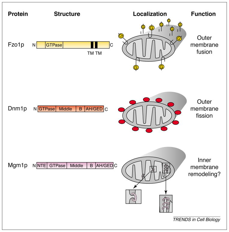
Mitochondria adopt a variety of different shapes in eukaryotic cells, ranging from multiple, small compartments to elaborate tubular networks. The establishment and maintenance of different mitochondrial morphologies depends, in part, on the equilibrium between opposing fission and fusion events. Recent studies in yeast, flies, worms and mammalian cells indicate that three high-molecular-weight GTPases control mitochondrial membrane dynamics. One of these is a dynamin-related GTPase that acts on the outer mitochondrial membrane to regulate fission. Recently, genetic approaches in budding yeast have identified additional components of the fission machinery. These and other new findings suggest a common mechanism for membrane fission events that has been conserved and adapted during eukaryotic evolution.


Figures

Fig. 1
Fig. 1
Yeast mitochondria form a dynamic reticulum. Wild-type yeast cells producing mitochondrion-targeted green fluorescent protein (mito-GFP) were grown to log phase in media containing galactose at 30°C A 3 μl aliquot was placed on a microscope slide under a coverslip and sealed with nail polish. (a) Stereo pair of a mito-GFP-labeled, unbudded yeast cell. Three-dimensional images were acquired using wide-field fluorescence microscopy. (b) Time-resolved images from a single optical section of 0.2 μm. Time increases from left to right in 3 min increments as indicated. Arrows on the 3 and 9 min images mark fusion events; the arrow on the 21 min image marks a fission event. Bar, 2 μm.
Fig. 2
Fig. 2
Three large GTPases control mitochondrial morphology in yeast. The predicted domain structures of Fzo1p, Dnm1p and Mgm1p, and their submitochondrial localization patterns are shown here. The known (Fzo1p, Dnm1p) or postulated (Mgm1p) function of each GTPase in mitochondrial membrane dynamics is indicated. The magnified views below the Mgm1p localization diagram show possible roles of Mgm1p in inner membrane fission (left) or cristae formation (right).
Fig. 3
Fig. 3
Dnm1p and Fzo1p act in opposing fission and fusion pathways to maintain the yeast mitochondrial network. Ongoing fission and fusion events maintain a tubular mitochondrial network in wild-type yeast cells (middle). Loss of the Dnm1p GTPase (dnm1Δ) leads to net formation owing to unopposed mitochondrial tip fusion (left). Loss of the Fzo1p GTPase (fzo1Δ) leads to fragmentation owing to unopposed mitochondrial fission (right).
Fig. 4
Fig. 4
Mdv1p and Fis1p regulate Dnm1p-mediated mitochondrial fission. (a) Predicted domain structures and mitochondrial localization patterns of Dnm1p, Mdv1p and Fis1p. (b) The dependence of Dnm1p’s mitochondrial localization pattern on FIS1 and MDV1 function. The localization pattern of Dnm1p (red circles) is shown on the outer mitochondrial membrane in (from left to right) wild-type cells, fis1 mutant cells, mdv1 mutant cells and mdv1 fis1 mutant cells. The fission phenotype observed is indicated below each cell type. (c) The dependence of Mdv1p’s mitochondrial localization pattern on DNM1 and FIS1 function. The localization pattern of Mdv1p (blue circles) is shown on the outer mitochondrial membrane in (from left to right) wild-type cells, fis1 mutant cells, dnm1 mutant cells and dnm1 fis1 mutant cells. In dnm1 fis1 mutant cells, Mdv1p localizes to the cytoplasm and is not shown (far right). The fission phenotype observed is indicated below each cell type. Abbreviations: AH, α-helical domain; NTE, N-terminal extension; TM, transmembrane domain.
Fig. 5
Fig. 5
Molecular model of mitochondrial division. T op: mitochondrial division is depicted as a linear pathway with the relative rates of each step (1–3) indicated. Red circles represent Dnm1p-containing complexes. Bottom: the postulated molecular interactions occurring between Dnm1p, Fis1p and Mdv1p during steps 1 and 2 of the pathway. Mitochondrial tubules are drawn in cross section and a single complex is shown containing the three molecules. Dnm1p, Mdv1p and Fis1p are shown in red, blue, and green, respectively. Abbreviation: IMS, intermembrane space.

References

    1. Frank S, et al. The role of dynamin-related protein 1, a mediator of mitochondrial fission, in apoptosis. Dev Cell. 2001;1:515–525. - PubMed
    1. Hermann GJ, Shaw JM. Mitochondrial dynamics in yeast. Annu Rev Cell Dev Biol. 1998;14:265–303. - PubMed
    1. Yaffe MP. The machinery of mitochondrial inheritance and behavior. Science. 1999;283:1493–1497. - PubMed
    1. Jensen RE, et al. Yeast mitochondrial dynamics: fusion, division, segregation and shape. Microsc Res Tech. 2001;51:573–583. - PubMed
    1. Johnson LV, et al. Localization of mitochondria in living cells with rhodamine 123. Proc Natl Acad Sci U S A. 1980;77:990–994. - PMC - PubMed

Publication types

MeSH terms

Substances

LinkOut - more resources