. 2002 May;86(3):F171-7.
doi: 10.1136/fn.86.3.f171.

Magnetic resonance imaging of the infant brain: anatomical characteristics and clinical significance of punctate lesions

Affiliations

Magnetic resonance imaging of the infant brain: anatomical characteristics and clinical significance of punctate lesions

L G Cornette et al. Arch Dis Child Fetal Neonatal Ed. 2002 May.

Abstract

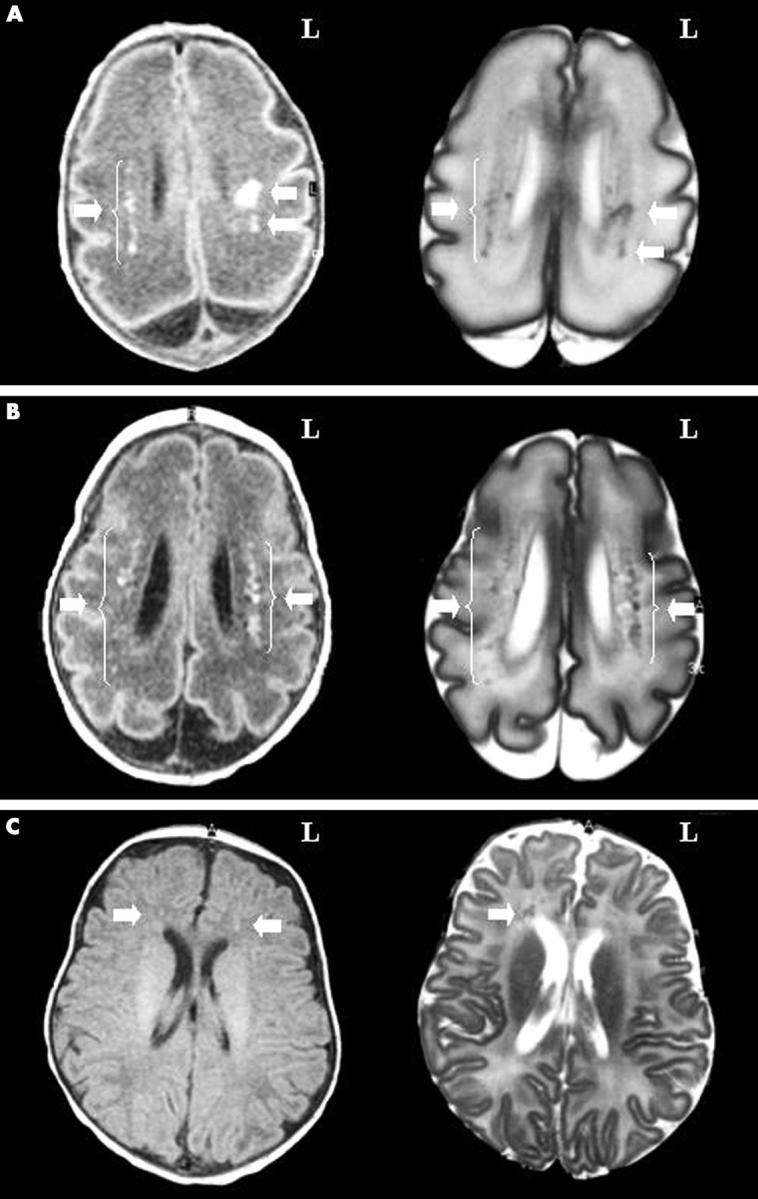
Objective: To describe the magnetic resonance imaging (MRI) characteristics of punctate brain lesions in neonates (number, appearance, distribution, and association with other brain abnormalities) and to relate them to neurodevelopmental outcome.

Methods: A retrospective analysis was performed of 110 MRI brain scans from 92 infants admitted in 1998 to the neonatal intensive care unit. Results of routine neurodevelopmental follow up (1998-2001) in those infants with punctate brain lesions were analysed.

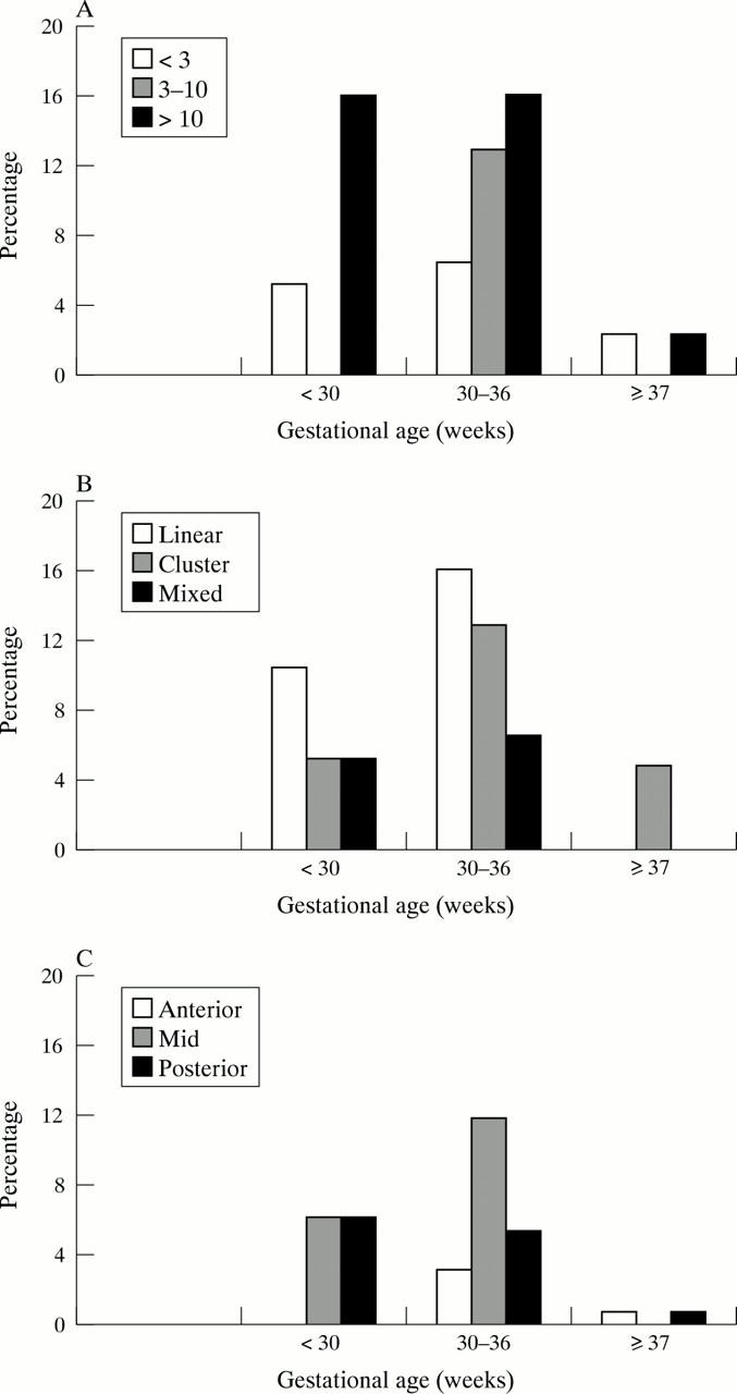
Results: Punctate lesions were observed in 15/50 preterm and 2/42 term infants. In the preterm group, the number of lesions was < 3 in 20%, 3-10 in 27%, and > 10 in 53%. In 14/15 the lesions were linearly organised and located in the centrum semiovale. Other brain abnormalities were absent or minor--that is, "isolated" punctate lesions--in 8/15 and major in 7/15. In the term group, punctate lesions were organised in clusters and no other brain abnormalities were observed. Isolated punctate lesions were observed in 10/17 infants, and a normal neurodevelopmental outcome was seen in 9/10 (mean follow up 29.5 months). One infant showed a slight delay in language development. In the infants with associated brain lesions (7/17, mean follow up 27.5 months), outcome was normal in only two subjects.

Conclusions: Punctate lesions are predominantly seen in preterm infants, are usually linearly organised, and border the lateral ventricles. Isolated punctate lesions may imply a good prognosis, because most of these subjects have a normal neurodevelopmental outcome so far.


Figures

Figure 1
Figure 1
Magnetic resonance characterisation of punctate lesions. (A) Isolated punctate lesions in an infant of 28 weeks gestation, scanned at 7 days of age (corresponding to case 1 in table 1). Left panel: T1 weighted axial image through the body of the lateral ventricles. Bilateral high intensity spots (arrows) correspond to punctate lesions, predominantly seen in the mid and posterior regions of the brain. In the right hemisphere, lesions (about 10) are linearly organised in the periventricular white matter. In the left hemisphere, lesions are organised in a mixed-type pattern (clustered and linear). No other brain abnormalities are observed. L, Left. Right panel: corresponding T2 weighted axial image of same infant. Bilateral low intensity spots (arrows) correspond to punctate lesions. (B) Punctate lesions in an infant of 29 weeks gestation with cystic periventricular leucomalacia (PVL), scanned at 26 days of age (corresponding to case 11 in table 1). Left panel: T1 weighted axial image through the body of the lateral ventricles. Bilateral high intensity spots (arrows) correspond to punctate lesions, predominantly seen in the mid and posterior regions of the brain. In the two hemispheres, the lesions are linearly organised and border a medially located discrete band of low signal intensity lesions in the periventricular white matter. Right panel: corresponding T2 weighted axial image of same infant. Bilateral low intensity spots (arrows) correspond to punctate lesions. The punctate lesions are clearly neighbouring a medial region of cystic PVL (high intensity lesions, corresponding to cystic degeneration). (C) Isolated punctate lesions in an infant of 40 weeks gestation, scanned at 51 days of age (corresponding to case 16 in table 1). Left panel: T1 weighted axial image through the body of the lateral ventricles. Bilateral high intensity spots (arrows) correspond to punctate lesions, exclusively seen in the anterior region of the brain. No anatomical or myelination abnormalities are observed. Right panel: corresponding T2 weighted axial image of same infant. Low intensity spots (arrow) correspond to punctate lesions. In the right hemisphere, a cluster of six lesions is observed, whereas only one spot is clearly distinguished on the T1 weighted image.
Figure 2
Figure 2
Overview of punctate lesions. Results are grouped according to the gestational age of all subjects analysed (n = 92): < 30 weeks (n = 19), between 30 and 36 weeks (n = 31), and ≥ 37 weeks (n = 42). (A) Number of punctate lesions. The percentage of cases with < 3, 3–10, or > 10 lesions is plotted for the different groups. (B) Appearance of punctate lesions. The percentage of cases with linear, clustered, or mixed appearance is plotted for the different groups. (C) Topography of punctate lesions. The percentage of cases in the anterior, mid, or posterior region of the brain is plotted for the different groups. As lesions were observed in the anterior, mid, and posterior region of the brain, we multiplied the total number of cases by three to calculate the percentage.

References

    1. Neuropediatrics. 1998 Aug;29(4):180-8 - PubMed
    1. Pediatrics. 1998 Aug;102(2 Pt 1):323-8 - PubMed
    1. AJNR Am J Neuroradiol. 1998 Nov-Dec;19(10):1909-21 - PubMed
    1. Pediatrics. 1999 Jan;103(1):39-46 - PubMed
    1. Neurology. 1999 Jul 13;53(1):132-9 - PubMed

Publication types