. 2002 May 1;22(9):3445-53.
doi: 10.1523/JNEUROSCI.22-09-03445.2002.

Neuronal deficiency of presenilin 1 inhibits amyloid plaque formation and corrects hippocampal long-term potentiation but not a cognitive defect of amyloid precursor protein [V717I] transgenic mice

Affiliations

Neuronal deficiency of presenilin 1 inhibits amyloid plaque formation and corrects hippocampal long-term potentiation but not a cognitive defect of amyloid precursor protein [V717I] transgenic mice

Ilse Dewachter et al. J Neurosci. .

Abstract

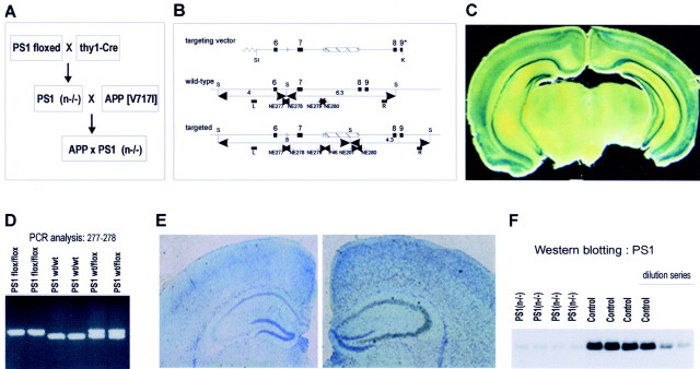
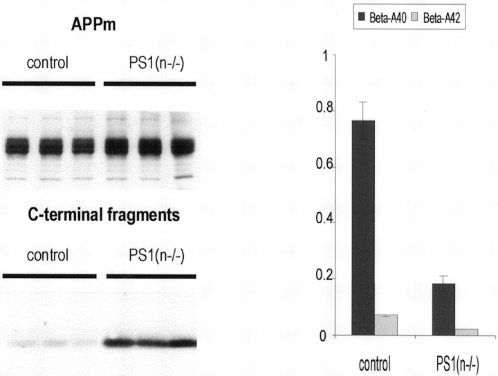
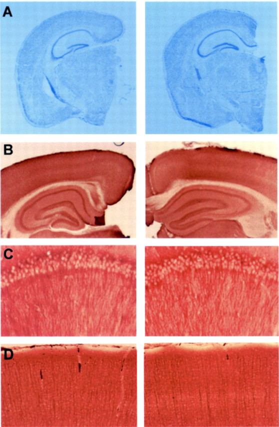
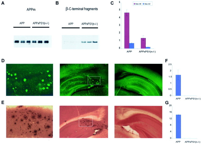
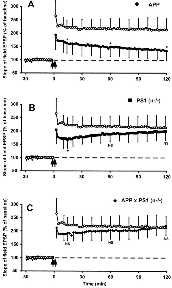
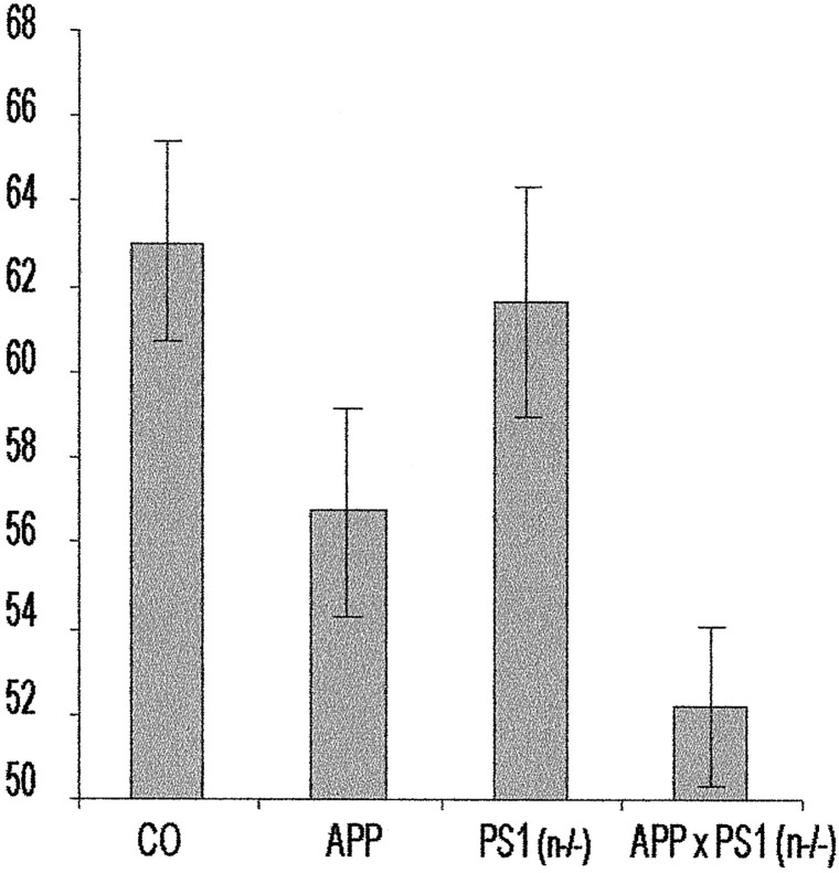
In the brain of Alzheimer's disease (AD) patients, neurotoxic amyloid peptides accumulate and are deposited as senile plaques. A major therapeutic strategy aims to decrease production of amyloid peptides by inhibition of gamma-secretase. Presenilins are polytopic transmembrane proteins that are essential for gamma-secretase activity during development and in amyloid production. By loxP/Cre-recombinase-mediated deletion, we generated mice with postnatal, neuron-specific presenilin-1 (PS1) deficiency, denoted PS1(n-/-), that were viable and fertile, with normal brain morphology. In adult PS1(n-/-) mice, levels of endogenous brain amyloid peptides were strongly decreased, concomitant with accumulation of amyloid precursor protein (APP) C-terminal fragments. In the cross of APP[V717I]xPS1 (n-/-) double transgenic mice, the neuronal absence of PS1 effectively prevented amyloid pathology, even in mice that were 18 months old. This contrasted sharply with APP[V717I] single transgenic mice that all develop amyloid pathology at the age of 10-12 months. In APP[V717I]xPS1 (n-/-) mice, long-term potentiation (LTP) was practically rescued at the end of the 2 hr observation period, again contrasting sharply with the strongly impaired LTP in APP[V717I] mice. The findings demonstrate the critical involvement of amyloid peptides in defective LTP in APP transgenic mice. Although these data open perspectives for therapy of AD by gamma-secretase inhibition, the neuronal absence of PS1 failed to rescue the cognitive defect, assessed by the object recognition test, of the parent APP[V717I] transgenic mice. This points to potentially detrimental effects of accumulating APP C99 fragments and demands further study of the consequences of inhibition of gamma-secretase activity. In addition, our data highlight the complex functional relation of APP and PS1 to cognition and neuronal plasticity in adult and aging brain.


Figures

Fig. 1.
Fig. 1.
Generation of PS1(n−/−) mice. A, Overall strategy to generate mice with a conditional inactivation of PS1, denoted PS1(n−/−), and APP transgenic mice with a neuronal PS1 deficiency. B, Schematic representation of the targeting vector, the wild-type, and the targeted PS1 gene. Lox P sites are represented by open triangles flanking the neomycin cassette and exon 7. Arrowheads represent synthetic primers used for genotyping by PCR. C, β-Galactosidase staining of brain section of the selected thy1-Cre-recombinase transgenic mouse line after crossing with the LacZ reporter mouse (Akagi et al., 1997) demonstrating neuronal staining in cortex and hippocampus. D, Genotyping by PCR analysis of DNA isolated from tail biopts using primers NE277 and NE278 (B) to identify the wild-type and targeted PS1 genes in mice with the indicated genotypes. E, In situ hybridization of brain sections of PS1(n−/−) mice (left panel) and of mice with a floxed but active PS1 gene (right panel) with RNA probes specific for PS1 exon 7, which is deleted by Cre-recombinase. F, Western blotting for the C-terminal fragment of mouse PS1 (∼20 kDa) in total brain extracts from four individual PS1(n−/−) mice [lanes marked PS1(n−/−)] and from four individual mice with floxed but active PS1 genes (lanes marked Control), and diluted sample (1, 0.5, 0.25). Quantitation revealed levels of 100.0 ± 5.5 and 16.0 ± 0.7% in control mice and PS1(n−/−) mice, respectively. Western blotting with antibodies against APP and LRP was used as a loading control.
Fig. 2.
Fig. 2.
APP processing in brain of PS1(n−/−) mice. Western blotting (left panel) of mouse brain extracts demonstrating very similar levels of membrane-bound APP (APPm; ∼110 kDa) (top) and accumulation of the APP C-terminal fragments (∼10–12 kDa) (bottom) in PS1(n−/−) mice. Results shown are from three individual PS1(n−/−) mice (three right lanes) and from three individual mice with floxed but active PS1 genes (control, three left lanes). Antiserum B10/4 was used that is specific for the C-terminal domain of human and mouse APP (Dewachter et al., 2000). Levels of soluble amyloid peptides (right panel) (nanograms per gram brain tissue) extracted from the brain of mice with a floxed but active PS1 gene (control) and from PS1(n−/−) mice, as measured with specific ELISA for murine Aβ40 (black bars) and Aβ42 (gray bars) (mean with SEM,n = 6 for each genotype).
Fig. 3.
Fig. 3.
Analysis of Notch signaling and PS2 expression. Northern blotting analysis of RNA extracted from brain of mice with floxed but active PS1 genes (lanes 1–3) and of PS1(n−/−) mice (lanes 4–6) demonstrates no changes of mRNA levels of Hes5 (top) and Dll1 (middle). Western blotting for the C-terminal fragment of mouse PS2 (∼20 kDa) (bottom), in total brain extracts from four individual PS1(n−/−) mice [lanesmarked PS1(n−/−)] and from four individual mice with floxed but active PS1 genes (lanes markedControl), and diluted sample (1, 0.5, 0.25). Quantitation revealed levels of 100 ± 2 and 127 ± 11% in control mice and PS1(n−/−) mice, respectively.
Fig. 4.
Fig. 4.
Morphological analysis of brain of PS1(n−/−) mice. A, Cresyl violet staining of paraffin-embedded sections from brain of mice with floxed but active PS1 genes (left panel) and from PS1(n−/−) mice (right panel), aged 6 months, demonstrating normal brain architecture in PS1(n−/−) mice. B, Immunohistochemical staining for GAP43 of vibratome sections from brain of mice with floxed but active PS1 genes (left panel) and PS1(n−/−) mice (right panel), demonstrating normal morphology of hippocampus.C, D, Higher magnification of neurons in hippocampal CA1 region (C) and cortex (D) after immunohistochemical staining for MAP2 of vibratome sections from brain of mice with floxed but active PS1 genes (left) and PS1(n−/−) mice (right).
Fig. 5.
Fig. 5.
Inhibition of amyloid peptides and pathology in brain of APPxPS1(n−/−) mice. A, B, Western blotting of membrane proteins extracted from brain of three individual APP[V717I] transgenic mice (lanes markedAPP) and from three individual APPxPS1(n−/−) transgenic mice. The Western blots were stained with monoclonal antibody 1G5 for full-length transgene membrane-bound APP (APPm) (∼110 kDa) (A) and with monoclonal antibody WO2 for human APP β-C-stubs (∼12 kDa) (B). C, Levels of soluble amyloid peptides (nanograms per gram brain tissue) in brain of APP and APPxPS1(n−/−) transgenic mice as measured with specific ELISA for human Aβ40 (red bars) and Aβ42 (blue bars) (mean with SEM, n = 6 for each genotype). D, E, Staining with thioflavinS (D) and immunostaining (E) of amyloid deposits in brain of APPxPS1(n−/−) (middle panels) and APP[V717I] transgenic mice (right panels). Note the absence of thioflavin-S staining and of immunoreaction in the subiculum of APPxPS1(n−/−) mice in contrast to the parent APP[V717I] transgenic mice. The left panels are a higher magnification of the areas selected in the middle panels. F,G, Amyloid load, expressed as relative surface area occupied by thioflavin-S-positive plaques (F) and of immune-positive plaques (G) in APP[V717I] and APPxPS1(n−/−) mice, all 16–18 months old (mean with SEM,n = 6).
Fig. 6.
Fig. 6.
Long-term potentiation in hippocampal slices of APP[V717I], PS1(n−/−), and APPxPS1(n−/−) transgenic mice. The slope of field Schaffer's EPSP recorded before and after tetanic stimulation of Schaffer's collaterals in brain sections from APP[V717I] (A), PS1(n−/−) (B), and APPxPS1(n−/−) (C) mice in each panel compared with mice with floxed but active PS1 genes (○). Each data point shown is the mean ± SD of results from six individual mice of each genotype. A statistically significant decrease was evident for the fEPSP in APP[V717I] mice (p < 0.05; indicated byasterisk in A), as described previously (Moechars et al., 1999; Schneider et al., 2001). The final LTP after 2 hr was not significantly different in PS1(n−/−) mice or in APPxPS1(n−/−) mice (NS; p > 0.05) (B, C). Note the initial decrease 15 min after tetanic stimulation that is borderline statistically significant in the PS1(n−/−) mice (see Discussion).
Fig. 7.
Fig. 7.
Analysis of cognition of wild-type, PS1(n−/−), APP[V717I], and APPxPS1(n−/−) transgenic mice in the object recognition task. In the novel-object recognition task, recognition memory is expressed as exploratory preference in the retention test. The recognition index =tB/(tA +tB) × 100, withA and B, respectively, representing the familiar and the novel object. Exploratory preference for control mice, i.e., mice with floxed but active PS1 genes (bar markedCO; n = 22), PS1(n−/−) mice (n = 17), APP[V717I] mice (n= 24), and APPxPS1(n−/−) (n = 18) transgenic mice. All mice were 3–6 months old. Retention was measured at 1 and 3 hr after training (mean ± SEM). At 3 hr after training, the APP[V717I] and APPxPS1(n−/−) mice were significantly impaired compared with control mice (respectively, p < 0.05, p < 0.001; determined by ANOVA analysis).

References

    1. Akagi K, Sandig V, Vooijs M, Van der Valk M, Giovannini M, Strauss M, Berns A. Cre-mediated somatic site-specific recombination in mice. Nucleic Acids Res. 1997;25:1766–1773. - PMC - PubMed
    1. Calhoun ME, Burgermeister P, Phinney AL, Stalder M, Tolnay M, Wiederhold KH, Abramowski D, Sturchler-Pierrat C, Sommer B, Staufenbiel M, Jucker M. Neuronal overexpression of mutant amyloid precursor protein results in prominent deposition of cerebrovascular amyloid. Proc Natl Acad Sci USA. 1999;96:14088–14093. - PMC - PubMed
    1. Chapman PF, White GL, Jones MW, Cooper-Blacketer D, Marshall VJ, Irizarry M, Younkin L, Good MA, Bliss TV, Hyman BT, Younkin SG, Hsiao KK. Impaired synaptic plasticity and learning in aged amyloid precursor protein transgenic mice. Nat Neurosci. 1999;2:271–276. - PubMed
    1. De Strooper B, Saftig P, Craessaerts K, Vanderstichele H, Guhde G, Annaert W, Von Figura K, Van Leuven F. Deficiency of presenilin-1 inhibits the normal cleavage of amyloid precursor protein. Nature. 1998;391:387–390. - PubMed
    1. De Strooper B, Annaert W, Cupers P, Saftig P, Craessaerts K, Mumm JS, Schroeter EH, Schrijvers V, Wolfe MS, Ray WJ, Goate A, Kopan R. A presenilin-1-dependent gamma-secretase-like protease mediates release of Notch intracellular domain. Nature. 1999;398:518–522. - PubMed

Publication types

MeSH terms

LinkOut - more resources