. 2002 May 1;22(9):3473-83.
doi: 10.1523/JNEUROSCI.22-09-03473.2002.

Lentiviral-mediated delivery of mutant huntingtin in the striatum of rats induces a selective neuropathology modulated by polyglutamine repeat size, huntingtin expression levels, and protein length

Affiliations

Lentiviral-mediated delivery of mutant huntingtin in the striatum of rats induces a selective neuropathology modulated by polyglutamine repeat size, huntingtin expression levels, and protein length

Luis Pereira de Almeida et al. J Neurosci. .

Abstract

A new strategy based on lentiviral-mediated delivery of mutant huntingtin (htt) was used to create a genetic model of Huntington's disease (HD) in rats and to assess the relative contribution of polyglutamine (CAG) repeat size, htt expression levels, and protein length on the onset and specificity of the pathology. Lentiviral vectors coding for the first 171, 853, and 1520 amino acids of wild-type (19 CAG) or mutant htt (44, 66, and 82 CAG) driven by either the phosphoglycerate kinase 1 (PGK) or the cytomegalovirus (CMV) promoters were injected in rat striatum. A progressive pathology characterized by sequential appearance of ubiquitinated htt aggregates, loss of dopamine- and cAMP-regulated phosphoprotein of 32 kDa staining, and cell death was observed over 6 months with mutant htt. Earlier onset and more severe pathology occurred with shorter fragments, longer CAG repeats, and higher expression levels. Interestingly, the aggregates were predominantly located in the nucleus of PGK-htt171-injected rats, whereas they were present in both the nucleus and processes of CMV-htt171-injected animals expressing lower transgene levels. Finally, a selective sparing of interneurons was observed in animals injected with vectors expressing mutant htt. These data demonstrate that lentiviral-mediated expression of mutant htt provides a robust in vivo genetic model for selective neural degeneration that will facilitate future studies on the pathogenesis of cell death and experimental therapeutics for HD.


Figures
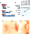
Fig. 1.
Fig. 1.
A, Schematic representation of the lentiviral constructs used in this study. cDNAs coding for the first 171, 853, and 1520 amino acids of human huntingtin with 19, 44, 66, or 82 CAG repeats were cloned in the SIN-W transfer vector.B, Western blot analysis, using the EM-48 antibody showing that huntingtin fragments of the expected molecular weights are produced in human embryonic kidney 293T cells infected with the corresponding lentiviral vectors. C–E, Photomicrographs of striatal sections immunostained with the Ab675 antibody recognizing the N-terminal part of huntingtin. One week after injection, both wild-type (htt171-19Q; C) and mutant (htt171-82Q;D) huntingtin fragments are overexpressed in a large area of the striatum. E, Sustained expression of the transgene was observed 12 weeks after injection.
Fig. 2.
Fig. 2.
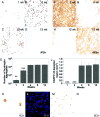
Time course analysis of the neuropathology in htt171-19Q- and htt171-82Q-infected rats. A–C, Huntingtin aggregates were first detected 1 week after injection of htt171-82Q and progressively accumulated during the 12 week study period. D, As expected, huntingtin inclusions were not identified with the EM48 antibody in the htt171-19Q-transduced hemisphere. E, Quantification of the number of cells containing huntingtin aggregates in the striatum of htt171-82Q-injected rats. **p < 0.01. F–H, Evaluation of the neurotoxicity of wild-type and mutant huntingtin fragments on DARPP-32-stained striatal sections. G, Four weeks after infection, a drastic loss of DARPP-32-immunoreactive neurons was observed in the htt171-82Q-infected striatum, whereas overexpression of the wild-type protein (htt171-19Q; I) had no deleterious effect on DARPP-32 staining even at 3 months.J, Quantification of the DARPP-32-depleted region on the htt171-82Q-transduced striatal neurons. *p < 0.05; **p < 0.01; ***p < 0.001.K, EM48 staining showing an immunoreactive nucleus with neuronal intranuclear inclusions. L, DAPI and EM48 double staining showing that most of the htt aggregates are located in the nucleus of htt171-82Q-infected neurons and colocalized with ubiquitin (M). N, Ubiquitin staining in htt171-19Q-injected rats.
Fig. 3.
Fig. 3.
Progressive striatal degeneration in htt171-82Q-injected rats. A, The first indication of striatal degeneration was observed at 4 weeks on Fluoro-Jade B-stained sections. C, At 8 weeks, pyknotic nuclei (arrowhead) were visible on cresyl violet-stained sections. E, Coalescence of the internal capsule of the striatum was observed at 12 weeks on a bright-field photomicrograph. In contrast, no signs of degeneration were observed with the wild-type huntingtin fragment (B, D, F, H). Finally, robust GFAP staining was observed 3 months after injection in mutant htt (G) compared with wild-type htt (H).
Fig. 4.
Fig. 4.
Specificity of the striatal toxicity in htt171-82Q-injected rats. Low-power (A) and high-power (B) photomicrographs of a striatal section triple-stained for DARPP-32 (red), ChAT (brown), and NADPH-d (blue) show that 12 weeks after the injection of htt171-82Q, ChAT-positive (arrowhead) and NADPH-d-positive (open triangle) interneurons are still present at the center of the DARPP-32 depleted region. Double staining shows that ChAT-positive (C, red) and NADPH-d-positive (E, blue) neurons are transduced and develop large neuronal intranuclear huntingtin aggregates (brown). D, Control, noninfected ChAT and NADPH-d neurons. F, Quantification of the data illustrating the selective sparing of ChAT and NADPH-d interneurons at 3 months.
Fig. 5.
Fig. 5.
Impact of huntingtin expression levels on the severity of the pathology. The DARPP-32-depleted areas significantly decreased in CMV-htt171-81Q-injected animals compared with PGK-htt171-82Q-injected rats. *p < 0.05; ***p < 0.001.
Fig. 6.
Fig. 6.
Impact of huntingtin expression levels on the formation of aggregates. We have previously shown that the CMV promoter leads to a reduced expression level in the rat brain compared with the PGK promoter. Decreasing the in vivo expression level of the Htt171-82Q fragment alters the subcellular localization of the huntingtin inclusions (A–D). Low-magnification (A) and high-magnification (B) photomicrographs show that the EM48-immunoreactive aggregates in PGK-htt171-82Q-injected animals are mainly restricted to the nucleus of infected neurons, whereas both nuclear and neuritic aggregates are observed in the CMV-htt171-82Q-injected animals (C, D).
Fig. 7.
Fig. 7.
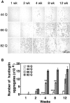
Impact of polyglutamine repeat size on the formation of huntingtin aggregates. A, Nuclear inclusions are first detected 4 weeks after injection of the htt171-44Q lentiviral vector and progressively accumulate over time. Increasing the CAG repeat size to 66 and 82 leads to an earlier appearance of aggregates and a significant increase in the number of nuclear inclusions. B, Quantification of the data showing the direct relationship between CAG repeat size and aggregate formation. *p < 0.05; **p < 0.01; ***p < 0.001.
Fig. 8.
Fig. 8.
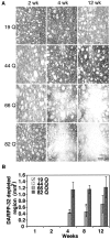
Influence of polyglutamine tract on the loss of DARPP-32 immunoreactivity. A, As expected, the overexpression of wild-type htt171 is not inducing a loss of DARPP-32 immunoreactivity. Interestingly, the DARPP-32 staining is also preserved in the htt171-44Q-injected animals, although huntingtin aggregates are first detected at 4 weeks in these animals. In the htt171-66Q-injected animals, the first indication of cellular dysfunction is observed at 4 weeks. Finally, a large DARPP-32-depleted region was present around the injection site in the htt171-82Q group.B, Quantification of the data showing the size of the DARPP-32-depleted region. *p < 0.05.
Fig. 9.
Fig. 9.
Impact of huntingtin protein length on the formation of aggregates. Increasing the huntingtin fragment size from 171 to 853 and 1520 amino acids significantly delays the appearance of neuronal aggregates. *p < 0.05; **p < 0.01; ***p < 0.001.
Fig. 10.
Fig. 10.
Striatal neuropathology 6 months after injection of lentiviral vectors coding for huntingtin fragments of various lengths. A, In htt171-82Q-injected rats, EM48-positive nuclear inclusions are still present but are mainly located at the limit of the degenerating area, whereas aggregates are detected around the injection site with the longer huntingtin fragments (C, E). Note that increasing the huntingtin fragment size modifies the subcellular localization of aggregates from nuclear to neuritic (A, C, E). B, D, F, The onset of the pathology, based on DARPP-32 immunoreactivity, is also significantly delayed with longer huntingtin fragments. A moderate loss of DARPP-32 staining is seen with the htt853 vector, whereas limited neuropathology, with DARPP-32 staining still present in neuronal cell bodies is observed in htt1520-82Q-injected rats.
Fig. 11.
Fig. 11.
Diagram summarizing the neuropathologies observed with the various huntingtin-expressing lentiviral vectors. Thearrows indicate the time points at which the initial pathological processes were observed based on huntingtin and DARPP-32 and NeuN immunoreactivity. The thickness of thearrows indicates the severity of the phenotype.

References

    1. Andrew SE, Goldberg YP, Kremer B, Telenius H, Theilmann J, Adam S, Starr E, Squitieri F, Lin B, Kalchman MA, Graham RK, Hayden MR. The relationship between trinucleotide (CAG) repeat length and clinical features of Huntington's disease. Nat Genet. 1993;4:398–403. - PubMed
    1. Andrews TC, Weeks RA, Turjanski N, Gunn RN, Watkins LH, Sahakian B, Hodges JR, Rosser AE, Wood NW, Brooks DJ. Huntington's disease progression. PET and clinical observations. Brain. 1999;122:2353–2363. - PubMed
    1. Antonini A, Leenders KL, Spiegel R, Meier D, Vontobel P, Weigell-Weber M, Sanchez-Pernaute R, de Yebenez JG, Boesiger P, Weindl A, Maguire RP. Striatal glucose metabolism and dopamine D2 receptor binding in asymptomatic gene carriers and patients with Huntington's disease. Brain. 1996;119:2085–2095. - PubMed
    1. Aylward EH, Brandt J, Codori AM, Mangus RS, Barta PE, Harris GJ. Reduced basal ganglia volume associated with the gene for Huntington's disease in asymptomatic at-risk persons. Neurology. 1994;44:823–828. - PubMed
    1. Aylward EH, Codori AM, Rosenblatt A, Sherr M, Brandt J, Stine OC, Barta PE, Pearlson GD, Ross CA. Rate of caudate atrophy in presymptomatic and symptomatic stages of Huntington's disease. Mov Disord. 2000;15:552–560. - PubMed

Publication types

MeSH terms