. 2002 May 1;22(9):3512-9.
doi: 10.1523/JNEUROSCI.22-09-03512.2002.

Muscle spindle-derived neurotrophin 3 regulates synaptic connectivity between muscle sensory and motor neurons

Affiliations

Muscle spindle-derived neurotrophin 3 regulates synaptic connectivity between muscle sensory and motor neurons

Hsiao-Huei Chen et al. J Neurosci. .

Abstract

Ia afferents induce the formation of muscle spindles prenatally and maintain them postnatally. To address whether spindles, in turn, regulate the function of Ia afferents, we examined Egr3-null mutant mice (Egr3-/-), in which muscle spindles degenerate progressively after birth. Egr3-/- mice develop gait ataxia, scoliosis, resting tremors, and ptosis, suggesting a defect in proprioception. Despite the normal morphological appearance of peripheral and central sensory projections, we observed a profound functional deficit in the strength of sensory-motor connections in Egr3-/- mice. Muscle spindles in Egr3-/- mice do not express NT3. Intramuscular injections of NT3 to Egr3-/- mice during the postnatal period restored sensory-motor connections. Thus, NT3 derived from muscle spindles regulates the synaptic connectivity between muscle sensory and motor neurons.


Figures

Fig. 1.
Fig. 1.
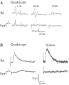
A, Method for measuring monosynaptic components of composite synaptic potentials. All threetraces show synaptic input from the obturator nerve recorded extracellularly from the L3 ventral root at P8. The monosynaptic model, shown in red, is scaled vertically to fit the rising phase of the synaptic potential. Terminal field potentials, indicated by open arrows, show the time at which Ia impulses arrive in the ventral horn. See Materials and Methods and Results for more details. Filled arrows inA and B indicate the beginning of the monosynaptic component. B, Monosynaptic EPSPs are severely reduced in Egr3−/− mice. Electrical stimulation of quadriceps, obturator, and soleus muscle nerves at P8 elicits EPSPs in motor axons recorded extracellularly from the ventral root. In normal mice [wild type (Wt)], the EPSPs consist of an early (∼5 msec latency), monosynaptic component from Ia fibers and a later, polysynaptic component from both Ia and Ib fibers. The early monosynaptic component is almost entirely missing in Egr3−/− mice, although the late polysynaptic component often remains.
Fig. 2.
Fig. 2.
GTO (Ib) afferents respond normally in Egr3−/− mice. A, A single muscle twitch evokes a burst of Ib spikes recorded from the dorsal root.B, Poststimulus time histograms of Ib activity (∼20 trials, shown as spikes/sec) show the similar time course of these responses in Egr3−/− and wild-type (Wt) mice. C, A burst of Ib activity evoked by a muscle twitch elicits a delayed synaptic response in motoneurons of both normal and mutant mice.
Fig. 3.
Fig. 3.
Central projections of sensory neurons appear anatomically normal in Egr3−/− mice at P8. Sensory axons including Ia afferents were labeled with HRP from the L3 dorsal root. There is a prominent projection, characteristic of Ia collateral axons, ventral to motoneurons in the lateral motor column at L3 in the vicinity of the quadriceps motor pool (A). Scale bar, 200 μm. B, At higher magnification of the region outlined in blue (inset inA), the terminal arborizations of Ia axons contain large numbers of swellings that appear to be synaptic boutons.
Fig. 4.
Fig. 4.
Ia afferents in Egr3−/− mice respond to low-frequency muscle stretch but fail to elicit monosynaptic EPSPs. A, At a 1 Hz repetition rate, small-amplitude passive muscle stretches evoke a burst of Ia spikes in both normal and mutant mice. When the stretches are applied at higher rates, however, spindles in mutant muscles fail to respond. B, Selective low frequency (0.5 Hz) stimulation of Ia afferents in quadriceps and soleus muscles elicits a subthreshold monosynaptic EPSP in motoneurons of wild-type (Wt) but not Egr3−/−mice.
Fig. 5.
Fig. 5.
Muscle spindles in Egr3−/− mice do not express NT3. A, B, Calbindin staining was used to identify spindles at P12. Spindles in Egr3−/− muscles are smaller and reduced in number compared with normal [wild-type (Wt)] mice. C, D, Mutant spindles at P12 are not immunopositive for NT3, unlike normal spindles. E, F, At P0, normal spindles express NT3 mRNA, but mutant spindles do not. Scale bar, 40 μm.
Fig. 6.
Fig. 6.
Synaptic connections in Egr3−/− mice are functional at birth but are lost by P2. Synaptic responses in motoneurons at P0 and P2 were evoked by electrical stimulation of the quadriceps muscle nerve and recorded in the L3 ventral root. Wt, Wild type.
Fig. 7.
Fig. 7.
Postnatal NT3 injections restore functional monosynaptic connections in Egr3−/−mice. A, Intramuscular NT3 injection during the first postnatal week at P1, P3, P5, and P7 [NT3(P1–7)] maintains functional synaptic connections. Representative records of synaptic responses in Egr3−/− mice are shown with or without NT3 injection relative to age-matched wild-type (Wt) mice. Arrows indicate the beginning of monosynaptic components. B, Analysis of EPSP amplitudes after NT3 injections. Four NT3 injections in the first postnatal week potentiated monosynaptic amplitude in wild-type mice (*p < 0.001) and in mutant mice (**p < 0.005). Three daily injections of NT3 between P5 and P7 [NT3(P5–7)] did not potentiate the monosynaptic response in wild-type mice (p = 0.55), nor did they restore monosynaptic responses in mutant mice (p = 0.18 vs mutants with no NT3 treatment). The number above each bar represents the numbers of cases examined.
Fig. 8.
Fig. 8.
A, B, Delayed injection of NT3 into Egr3−/− mice during the second postnatal week at P5, P7, P9, and P11 [NT3(P5–11)] restores synaptic connections. Monosynaptic responses in Egr3−/− mice with delayed NT3 injections are as large as in normal mice but are not potentiated relative to age-matched wild-type (Wt) mice (p = 0.54). Filled arrows indicate the beginning of monosynaptic components. Gray bars, Wild type;white bars, Egr3−/−.C, Delayed NT3 treatment does not fully restore conduction velocities in sensory axons. Traces are dorsal root recordings of quadriceps sensory axons at P12. Some axons in NT3-treated mutant mice conduct as rapidly as normal axons (presumably Ib axons), but others have an additional conduction delay of 1–2 msec. D, The terminal field potentials of Ia axons are also delayed (open arrows), resulting in an increase in the latency of the monosynaptic response.Traces are ventral root recordings of quadriceps input at P12.

References

    1. Arber S, Ladle DR, Lin JH, Frank E, Jessell TM. ETS gene Er81 controls the formation of functional connections between group Ia sensory afferents and motor neurons. Cell. 2000;101:485–498. - PubMed
    1. Arvanov VL, Seebach BS, Mendell LM. NT-3 evokes an LTP-like facilitation of AMPA/kainate receptor-mediated synaptic transmission in the neonatal rat spinal cord. J Neurophysiol. 2000;84:752–758. - PubMed
    1. Blasi J, Chapman ER, Link E, Binz T, Yamasaki S, De Camilli P, Sudhof TC, Niemann H, Jahn R. Botulinum neurotoxin A selectively cleaves the synaptic protein SNAP-25. Nature. 1993;365:160–163. - PubMed
    1. Brown MC, Engberg I, Matthews PB. The relative sensitivity to vibration of muscle receptors of the cat. J Physiol (Lond) 1967;192:773–800. - PMC - PubMed
    1. Carr VM, Simpson SB., Jr Proliferative and degenerative events in the early development of chick dorsal root ganglia. I. Normal development. J Comp Neurol. 1978;182:727–739. - PubMed

Publication types

MeSH terms