. 2002 May 1;22(9):3543-52.
doi: 10.1523/JNEUROSCI.22-09-03543.2002.

GAP-43 is critical for normal development of the serotonergic innervation in forebrain

Affiliations

GAP-43 is critical for normal development of the serotonergic innervation in forebrain

Stacy L Donovan et al. J Neurosci. .

Abstract

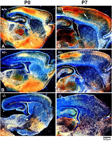
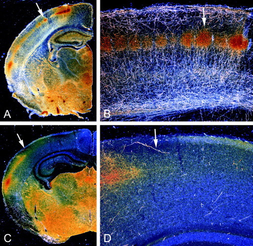
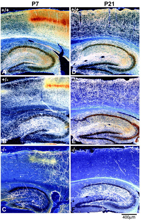
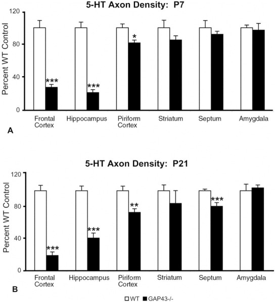
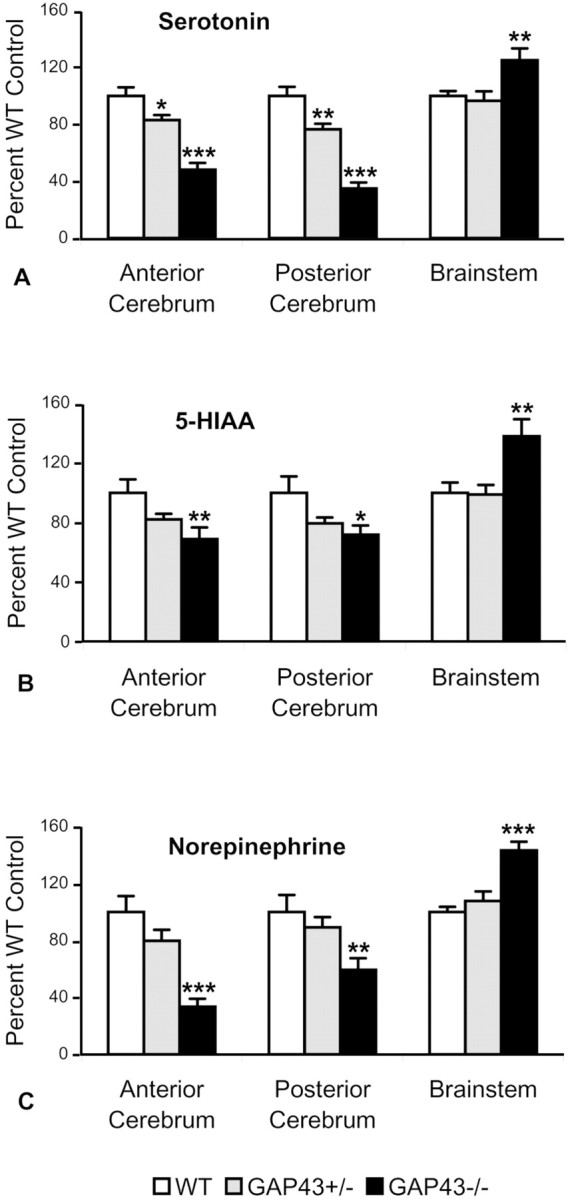
Serotonergic (5-HT) axons from the raphe nuclei are among the earliest afferents to innervate the developing forebrain. The present study examined whether GAP-43, a growth-associated protein expressed on growing 5-HT axons, is necessary for normal 5-HT axonal outgrowth and terminal arborization during the perinatal period. We found a nearly complete failure of 5-HT immunoreactive axons to innervate the cortex and hippocampus in GAP-43-null (GAP43-/-) mice. Abnormal ingrowth of 5-HT axons was apparent on postnatal day 0 (P0); quantitative analysis of P7 brains revealed significant reductions in the density of 5-HT axons in the cortex and hippocampus of GAP43-/- mice relative to wild-type (WT) controls. In contrast, 5-HT axon density was normal in the striatum, septum, and amygdala and dramatically higher than normal in the thalamus of GAP43-/- mice. Concentrations of serotonin and its metabolite, 5-hydroxyindolacetic acid, and norepinephrine were decreased markedly in the anterior and posterior cerebrum but increased in the brainstem of GAP43-/- mice. Cell loss could not account for these abnormalities, because unbiased stereological analysis showed no significant difference in the number of 5-HT dorsal raphe neurons in P7 GAP43-/- versus WT mice. The aberrant 5-HT innervation pattern persisted at P21, indicating a long-term alteration of 5-HT projections to forebrain in the absence of GAP-43. In heterozygotes, the density and morphology of 5-HT axons was intermediate between WT and homozygous GAP43-/- mice. These results suggest that GAP-43 is a key regulator in normal pathfinding and arborization of 5-HT axons during early brain development.


Figures

Fig. 1.
Fig. 1.
5-HT axons fail to innervate the cortex and hippocampus in neonatal GAP43−/− mice. Low-magnification dark-field photomicrographs of parasagittal sections from P0 (A–C) and P7 (D–F) mice show bright serotonin-immunoreactive axons on a dark background. At P0, 5-HT axons in WT mice are present throughout the brain and densely innervate the thalamus, basal forebrain, and frontal and parietal areas of the cortex (A). The density of 5-HT axons in GAP43−/− mice is dramatically reduced in most brain regions compared with WT mice at P0 (C). The density of 5-HT axons increases in the thalamus, cortex, and hippocampus of WT mice over the next week. At P7, distinct patches of serotonin immunoreactivity are visible in layer IV of the parietal cortex (arrow,D). In GAP43−/− mice at P7, the density of 5-HT axons in subcortical regions is similar to that in WT mice, but very few 5-HT axons are observed in the cortex and hippocampus (F). In GAP43+/− mice at P0 (B) and P7 (E), 5-HT axon density in the hippocampus and cortex is intermediate between that in GAP43−/− and WT mice. Scale bar, 500 μm.
Fig. 2.
Fig. 2.
The outgrowth of 5-HT axons to the dorsal neocortex and hippocampus falters in the absence of GAP-43. Dark-field photomicrographs show the distribution of 5-HT axons in coronal sections from P7 mice; arrows demarcate the same regions in low- and high-magnification views. In WT mice (A, B), 5-HT axons are present throughout the brain and densely innervate the thalamus, basal forebrain, hippocampus, and cortex. Distinct patches of serotonin immunoreactivity that demarcate barrels in layer IV of the parietal cortex are visible (arrows in A, B). In GAP43−/− mice (C, D), the growth of 5-HT axons into subcortical regions is relatively normal, but only a few 5-HT axons are observed beyond the ventral aspects of the neocortex.
Fig. 3.
Fig. 3.
The cortex and hippocampus exhibit persistent deficits in 5-HT innervation in GAP43−/− and GAP43+/− mice. Dark-field photomicrographs of parasagittal sections show 5-HT (A–C) and SERT (D–F) immunostaining in the cortex and hippocampus of P7 (A–C) and P21 (D–F) mice. At P7, 5-HT axons densely innervate the cortex and hippocampus in WT mice (A). 5-HT immunoreactivity is especially dense in layer IV of the primary somatosensory cortex, where barrel-like patches are clearly visible (A, right half). 5-HT axon density in layer IV of the primary somatosensory cortex of GAP43+/− mice at P7 (B) is similar to that in WT animals, but other cortical layers and the hippocampus show reduced densities of 5-HT axons. Only a few 5-HT axons are present in the hippocampus and cortex of P7 GAP43−/− mice (C). At P21, 5-HT axons labeled with SERT show a dense and even distribution throughout all layers of the cortex and hippocampus in WT mice (D). 5-HT innervation density in the cortex and hippocampus of GAP43+/− mice is less than that of WT mice at P21 (E). The density of 5-HT axons in the cortex remains extremely low in GAP43−/− mice at P21 (F). Scale bar, 400 μm.
Fig. 4.
Fig. 4.
5-HT axon densitometric analysis at P7 (A) and P21 (B) shows deficits in some but not all forebrain regions. Values are expressed as a percentage of the WT control mean. The density of 5-HT axons in the frontal cortex, hippocampus, and piriform cortex of GAP43−/− mice (black bars) is significantly reduced compared with WT mice (white bars) at both ages examined. In contrast, subcortical regions such as the amygdala and striatum show no significant differences in 5-HT innervation densities at both ages. 5-HT axon density in the septum is significantly reduced compared with WT at P21 but not at P7. *p ≤ 0.05; **p ≤ 0.01; ***p ≤ 0.001.
Fig. 5.
Fig. 5.
Selected brain regions from P7 mice show gene-dose-dependent effects on levels of serotonin, 5-HIAA, and norepinephrine as measured by HPLC. Values are expressed as percentage of WT controls. Serotonin levels are reduced by 52% in the anterior cerebrum and 65% in the posterior cerebrum and increased by 25% in the brainstem of GAP43−/− mice (black bars) compared with WT controls (white bars). GAP43+/− mice (gray bars) show decreasing levels of serotonin in the anterior (17%) and posterior (23%) cerebrum (A). Similar trends are observed for 5-HIAA levels. In GAP43−/− mice, 5-HIAA levels are reduced by 31 and 28% in the anterior and posterior cerebrum, respectively, whereas they increased by 38% in the brainstem (B). Levels of norepinephrine also decrease by 66 and 41% in the anterior and posterior cerebrum, respectively, and increase by 43% in the brainstem (C). *p < 0.05; **p < 0.01; ***p < 0.001.
Fig. 6.
Fig. 6.
Alterations in the density and morphology of 5-HT axons persist in the frontal cortex at P21. High-magnification dark-field and bright-field photomicrographs show the pattern and morphology of 5-HT axons in GAP43−/− mice at P21. A fine, dense network of 5-HT axons innervates all cortical layers of the frontal cortex in WT mice (A). A gene-dose-dependent reduction in the density of 5-HT axons is observed in GAP43+/− mice (B) and GAP-43–null mice (C). The morphology of 5-HT axons also varies according to genotype (D, E). In WT mice, 5-HT axons are fine and highly branched (A, D). In GAP43−/− mice (C, E), 5-HT axons show less branching and are coarser than in WT animals. The morphology of 5-HT axons in GAP43+/− mice is intermediate between that in WT and GAP43−/− mice (B). Scale bars: A–C, 200 μm;D, E, 10 μm.
Fig. 7.
Fig. 7.
At P7 and P21, 5-HT axons hyperinnervate the VB thalamus in GAP43−/− mice. Dark-field photomicrographs of coronal sections show 5-HT immunostaining in the thalamus of P7 (A–C) and P21 (D–F) mice. At P7, 5-HT axon density is dramatically higher in the VB thalamus of GAP43−/− mice (C) than in WT mice (A) and GAP43+/− mice (B). At P21, 5-HT axon density remains elevated in GAP43−/− mice (F) relative to GAP43+/− mice (E) and WT mice (D). Interestingly, 5-HT immunostaining of barreloid patches (arrows) and thalamocortical axons (asterisks) observed in WT (A) and heterozygote (B) animals at P7 is largely absent in GAP43−/− mice (C). Scale bar, 200 μm.
Fig. 8.
Fig. 8.
Densitometric analysis of the VB thalamus at P7 and P21 shows persistent increases in 5-HT axon density in GAP43−/− mice. Values are expressed as a percentage of the WT control mean. The density of 5-HT axons in the VB thalamus of GAP43−/− mice (white bars; 427%) and GAP43+/− mice (gray bars; 158%) is significantly increased compared with WT mice (black bars; 100%) at P7. 5-HT axon density remains elevated in GAP43−/− mice (158%) compared with WT and heterozygote mice at P21. *p ≤ 0.05, ***p ≤ 0.001 relative to WT;+p ≤ 0.05,++p ≤ 0.01 for GAP43+/− versus GAP43−/− comparisons; not significant, GAP43+/− versus WT at P21.

References

    1. Alonso G, Ridet JL, Oestreicher AB, Gispen WH, Privat A. B-50 (GAP-43) immunoreactivity is rarely detected within intact catecholaminergic and serotonergic axons innervating the brain and spinal cord of the adult rat, but is associated with these axons following lesion. Exp Neurol. 1995;134:35–48. - PubMed
    1. Andrews AM, Ladenheim B, Epstein CJ, Cadet JL, Murphy DL. Transgenic mice with high levels of superoxide dismutase activity are protected from the neurotoxic effects of 2′-NH2-MPTP on serotonergic and noradrenergic nerve terminals. Mol Pharmacol. 1996;50:1511–1519. - PubMed
    1. Azmitia EC, Dolan K, Whitaker-Azmitia PM. S-100β but not NGF, EGF, insulin or calmodulin is a CNS serotonergic growth factor. Brain Res. 1990;516:354–356. - PubMed
    1. Baumgarten HG, Grozdanovic Z. Psychopharmacology of central serotonergic systems. Pharmacopsychiatry. 1995;28 [Suppl 2]:73–79. - PubMed
    1. Bendotti C, Servadio A, Samanin R. Distribution of GAP-43 mRNA in the brain stem of adult rats as evidenced by in situ hybridization: localization within monoaminergic neurons. J Neurosci. 1991;11:600–607. - PMC - PubMed

Publication types

MeSH terms

LinkOut - more resources