. 2002 May 1;22(9):3568-79.
doi: 10.1523/JNEUROSCI.22-09-03568.2002.

Deleted in colorectal carcinoma and differentially expressed integrins mediate the directional migration of neural precursors in the rostral migratory stream

Affiliations

Deleted in colorectal carcinoma and differentially expressed integrins mediate the directional migration of neural precursors in the rostral migratory stream

Shin-ichi Murase et al. J Neurosci. .

Abstract

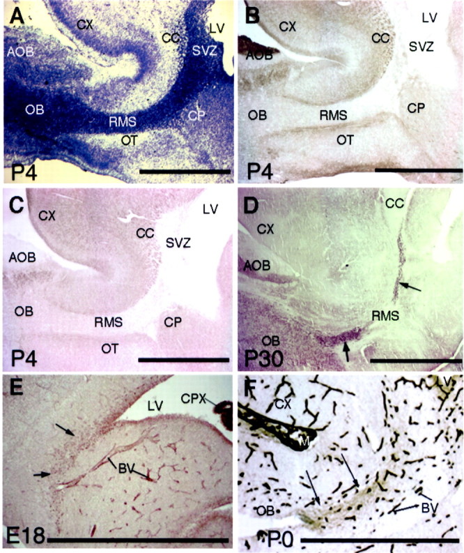
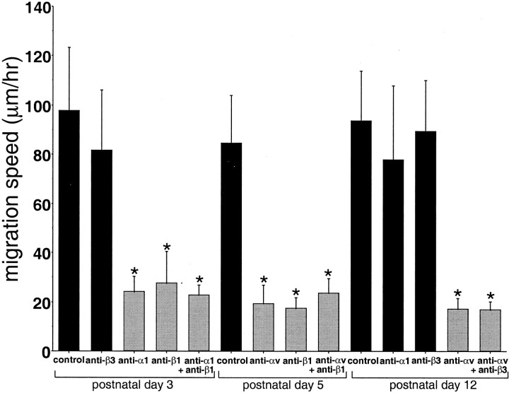
Precursors of the olfactory interneurons migrate from the subventricular zone via the rostral migratory stream (RMS). To investigate the molecular mechanisms by which RMS cells migrate, we used a slice preparation, which allows the migrating cells to be imaged at very high temporal and spatial resolution in the presence of added inhibitors. Using immunohistochemistry, we first determined that the alpha1-, beta8-, and beta1-integrin subunits and the alpha5- and gamma1-laminin subunits are expressed during embryonic day 16 to the early postnatal stage. During early postnatal days, alpha(v)- and beta6-integrins appeared, and their expression persisted throughout adulthood. The migrating cells also expressed the netrin receptors neogenin and Deleted in Colorectal Carcinoma (DCC). Netrin-1 is expressed in olfactory mitral cells. Anti-integrin antibodies inhibited the production of protrusions as well as cellular translocation. In contrast, anti-DCC antibodies primarily altered the direction of the protrusions; consequently, the migration was no longer unidirectional, and the speed was reduced. Thus, the interaction of DCC, possibly through an interaction with netrin-1, contributes to the direction of migration by regulating the formation of directed protrusions. In contrast, the integrins function in production of protrusions and cellular translocation, with different integrins participating at different developmental stages.


Figures

Fig. 1.
Fig. 1.
Experimental design for assays of cell migration in the RMS. Two hundred-micrometer-thick sagittal slices were cut from brains dissected from embryonic day 18 or postnatal day 0–16 mice using a vibratome. Slices containing the entire RMS were selected and placed on Millicell inserts in CCM1 medium containing HEPES and 5% horse serum. Crystals of DiI were placed on the RMS using a microneedle. By selecting the position of the DiI crystals, migrating cells in every part of the RMS can be visualized. Time-lapse images of fluorescent cells were recorded over 3–10 hr using an inverted microscope.
Fig. 2.
Fig. 2.
N-CAM, PSA, and laminin expression in the rostral migratory stream. A, Nissl staining of a sagittal section from a postnatal day 4 rat forebrain. The RMS, which is characterized by a high cellular density, begins at the anterior portion of the SVZ and ends at the center of the olfactory bulb (OB). B, C, N-CAM and PSA in the RMS of early postnates. B and C are adjacent sections from a P4 rat forebrain that are stained with anti-N-CAM (clone 5B8) and anti-PSA (clone MenB) MABs, respectively. The SVZ and the RMS stain very weakly, whereas the borders of the RMS show strong staining. D, PSA immunostaining of a P30 mouse forebrain. In contrast to weak expression of PSA in P4 RMS shown inC, PSA is expressed strongly in the RMS (arrows) and the olfactory bulb (OB).E, α5 subunit of laminin in the RMS (arrows) from an embryonic day 18 mouse shows punctate staining. Relatively strong staining is also seen in the choroid plexus (CPX) of the lateral ventricle (LV) and blood vessels (BV). F, γ1-Laminin is expressed in the RMS (arrows) from a P0 rat brain. Blood vessels (BV) and the meninges (M) are also stained. Scale bars:A–D, F, 1 mm; E, 50 μm.AOB, Accessory olfactory bulb; CC, corpus callosum; CP, caudate-putamen; CX, cerebral cortex; OT, olfactory tubercle.
Fig. 3.
Fig. 3.
Six integrins are differentially expressed in the rostral migratory stream. The α1 (A) and β8 (B) integrin subunits are expressed (arrows) from the anterior horn of the subventricular zone to the center of the olfactory bulb from P0 mice.C, The β1-integrin subunit is expressed in the RMS (arrows), blood vessels (BV), and the choroid plexus (CPX) of the lateral ventricle (LV) of a P2 mouse. D, The αv-integrin subunit is found in the RMS (arrows) from a P30 mouse. E, The β6-integrin subunit is expressed in the RMS (arrows) of a P15 mouse. F, The β3-integrin subunit is observed in P30 rat RMS (arrows). Scale bar, 1 mm. AOB, Accessory olfactory bulb; CC, corpus callosum; CX, cerebral cortex.
Fig. 4.
Fig. 4.
Summary of the stage-specific expression of integrin and laminin subunits in the RMS. Tenascin-C is expressed along the sides of the RMS but not in the RMS itself.
Fig. 5.
Fig. 5.
Integrins mediate migration of neural precursors in the RMS. Brain slices from a P12 mouse were labeled with DiI and cultured in CCM1 medium with 5% horse serum for 5 hr in the presence of either control or anti-integrin antibodies. Three hours after addition of DiI, the migration was confirmed by fluorescence time lapse, and then a control or function-blocking anti-αv-integrin antibody was added for (Figure legend continued.) 5 hr, after which migration was recorded over the next 3 hr. A, Time-lapse sequence of three cells (a–c) in a slice migrating from the SVZ (bottom) toward the olfactory bulb (top) in the presence of a control antibody. Thearrow pointing to each cell shows the leading process, and the line shows the cell body. The interval between each image is 5 min. See Movie 1 (available at www.jneurosci.org).B, Graphical representation of the migration of the three cells (a–c) in A. Eachpoint represents the position of the cell body at 5 min time points. Note the unidirectional pathway and the bursts of rapid migration followed by slower meandering. C, Time-lapse sequence of images of seven cells (d–j) in a slice migrating from the SVZ (left) to the olfactory bulb (right) in the presence of an anti-αv-integrin antibody. Seven cells (d–j) are marked for reference. See Movie 2 (available at www.jneurosci.org). D, Graphical representation of the migration of the seven cells as described in C. The olfactory bulb is at theright. Note the inhibited migration. The interval between each image is 5 min. Scale bars, 50 μm.
Fig. 6.
Fig. 6.
Inhibited migration of RMS cells by function-blocking anti-integrin antibodies. Living brain slices were prepared from P3, P5, and P12 mice, and the cells were labeled with a small crystal of DiI placed on the center of the RMS. The slices were cultured in CCM1 medium supplemented with HEPES and 5% horse serum. The slices were preincubated with anti-integrin antibodies for 5 hr, and the migrating cells were traced by time-lapse recording for 3 hr. At P3, when α1- and β1-integrins are expressed, corresponding blocking antibodies reduced the migration speed. Anti-β3 antibody did not inhibit the migration significantly. At P5, when αv- and β1-integrins are expressed, antibodies against these integrins inhibited the migration speed. At P12, when αv-integrin is expressed, anti-αv-integrin antibody inhibited the speed as well; however, anti-α1 and -β3 antibodies did not inhibit the migration speed significantly, and they are not expressed at this stage. Each value represents the mean ± SD. Statistical analysis was performed by one-way ANOVA with Scheffé's multiple comparison procedure (significance of p < 0.01). The groups withasterisks do not differ from each other, nor do the nonmarked groups, but in all other comparisons, the differences are significant.
Fig. 7.
Fig. 7.
Expression of DCC, neogenin, and netrin-1 and function of DCC in RMS migrations. A, Neogenin, a netrin-1 receptor, immunoreactivity coincides with the contour of RMS beginning at the anterior horn of the lateral ventricle (LV) and ending at the center of the olfactory bulb in a P0 mouse. B, The rostral part of the RMS strongly expresses the DCC protein, which is also present in the lateral olfactory tract (LOT) in P2 rats.AOB, Accessory olfactory bulb. C, Netrin-1 protein is expressed in the basal portion of olfactory mitral cells in the mitral cell layer (MCL) from an embryonic day 18 mouse. EPL, External plexiform layer;GCL, granule cell layer; GL, glomerular layer; ONL, olfactory nerve layer. (Figure legend continued.) D, Preabsorbed netrin-1 antibody (control) was prepared by coincubation of antibody and antigen peptide. The absorbed antibody did not show immunoreactivity. E, Sequence of 21 time-lapse images of a living slice from a P3 mouse treated with anti-DCC antibody as described in the legend to Figure 5. The olfactory bulb is located at the top. Three migrating cells (a–c) are indicated. Arrows indicate retracting leading process, and crossed arrows indicate processes pointing toward the anterior region of the subventricular zone (bottom). Note that the migration is no longer unidirectional, and that the processes form and retract frequently, which also distinguishes these migrations from those of normal cells shown in Figure 5. The interval between each image is 10 min. See Movie 3 (available atwww.jneurosci.org). F, Graphical representation of three migrating cells (a–c) shown in E. Cell bodies are tracked as described in Figure 5, and the time interval is 5 min. S, Start point; E, end point for cell b. See Movie 3 (available at www.jneurosci.org).G, Inhibited migration of cells by anti-DCC function-blocking antibodies. Living brain slices were prepared from P3 mice, and then the cells were labeled with DiI placed at the center or end of the RMS. The slices were cultured in CCM1 medium with HEPES and 5% horse serum with or without anti-DCC antibody for 5 hr, and the migration was observed by time-lapse recording. Each value represents the mean ± 1 SD. Statistical analysis was performed by one-way ANOVA with Scheffé's multiple comparison procedure (significance of p < 0.01). The control groups without anti-DCC antibody differ from every other group with anti-DCC. The groups withasterisks do not differ from each other, nor do the nonmarked groups, but in all other comparisons, the differences are significant. In contrast to cell treated with anti-integrin antibodies (Fig. 6), these cells show a larger net translocation. Scale bars:A, B, 1 mm; C, D, 100 μm;E, 50 μm.
Fig. 8.
Fig. 8.
Diagram depicting a working hypothesis for the roles of DCC and netrin and integrin in the migration of neural precursors from the SVZ to the center of the olfactory bulb. Netrin-1 secreted from mitral cells attracts DCC- or neogenin-expressing migrating cells, or both, to the olfactory bulb. Slit proteins from the septum inhibit migration out of the RMS into the septum or surrounding tissues by their repulsive activity. Integrins and laminins provide the traction for the motive force, and PSA-N-CAM provides the cellular milieu where the cells can move easily and maintenance of chains. Theshort arrow indicates the differentiation of neural precursors to granule cells; the long arrow marks the periglomerular cells. EPL, External plexiform layer;GCL, granule cell layer; GL, glomerular layer; MCL, mitral cell layer; ONL, olfactory nerve layer.

Similar articles

Cited by

References

    1. Allen E. The cessation of mitosis in the central nervous system of the albino rat. J Comp Neurol. 1912;22:547–568.
    1. Altman J. Autoradiographic and histological studies of postnatal neurogenesis. IV. Cell proliferation and migration in the anterior forebrain, with special reference to persisting neurogenesis in the olfactory bulb. J Comp Neurol. 1969;137:433–457. - PubMed
    1. Aufderheide E, Ekblom P. Tenascin during gut development: appearance in the mesenchyme, shift in molecular forms, and dependence on epithelial-mesenchymal interactions. J Cell Biol. 1988;107:2341–2349. - PMC - PubMed
    1. Boulder Committee: Angevine JBJ, Bodian D, Coulombre AJ, Edds MVJ, Hamburger V, Jacobson M, Lyser KM, Prestige MC, Sidman RL, Varon S, Weiss PA. Embryonic vertebrate central nervous system: revised terminology. Anat Rec. 1970;166:257–261. - PubMed
    1. Bryans WA. Mitotic activity in the brain of the adult rat. Anat Rec. 1959;133:65–73.

Publication types

MeSH terms