Clinical Trial
. 2002 May 1;22(9):3708-19.
doi: 10.1523/JNEUROSCI.22-09-03708.2002.

Prefrontal dopamine D1 receptors and working memory in schizophrenia

Affiliations
Clinical Trial

Prefrontal dopamine D1 receptors and working memory in schizophrenia

Anissa Abi-Dargham et al. J Neurosci. .

Abstract

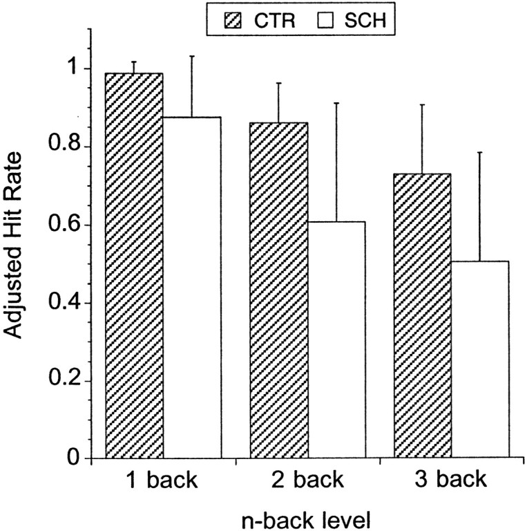
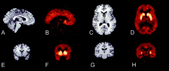
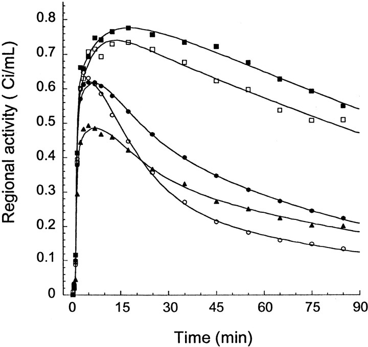
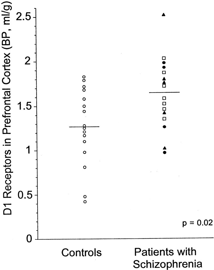
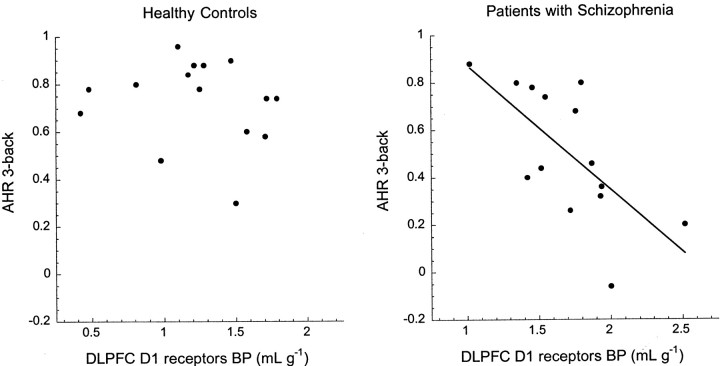
Studies in nonhuman primates documented that appropriate stimulation of dopamine (DA) D1 receptors in the dorsolateral prefrontal cortex (DLPFC) is critical for working memory processing. The defective ability of patients with schizophrenia at working memory tasks is a core feature of this illness. It has been postulated that this impairment relates to a deficiency in mesocortical DA function. In this study, D1 receptor availability was measured with positron emission tomography and the selective D1 receptor antagonist [11C]NNC 112 in 16 patients with schizophrenia (seven drug-naive and nine drug-free patients) and 16 matched healthy controls. [11C]NNC 112 binding potential (BP) was significantly elevated in the DLPFC of patients with schizophrenia (1.63 +/- 0.39 ml/gm) compared with control subjects (1.27 +/- 0.44 ml/gm; p = 0.02). In patients with schizophrenia, increased DLPFC [11C]NNC 112 BP was a strong predictor of poor performance at the n-back task, a test of working memory. These findings confirm that alteration of DLPFC D1 receptor transmission is involved in working memory deficits presented by patients with schizophrenia. Increased D1 receptor availability observed in patients with schizophrenia might represent a compensatory (but ineffective) upregulation secondary to sustained deficiency in mesocortical DA function.


Figures

Fig. 1.
Fig. 1.
Illustration of the steps involved in the sampling of activity from cortical regions. A coronal section 0.5 mm anterior to the rostral part of the corpus callosum is displayed. The MRI (A) is segmented into white matter, gray matter, and CSF voxels. Gray matter voxels are assigned a value of 1, and all other pixels are assigned a value of 0 to form a gray inverted mask image (B). The coregistered PET image (C) is multiplied by the mask (B) to form a gray matter PET image (D). Regional activities are sampled onD in nonzero voxels. Region boundaries (white lines) are illustrated on the right. Theyellow line corresponds to the AC–PC plane. The region ventral to this plane is the OFC. Regions dorsal to the AC–PC plane are divided into a lateral region (DLPFC) and medial regions, which, at this level, include the MPFC and ACC, dorsal and ventral to the cingulate gyrus, respectively.
Fig. 2.
Fig. 2.
Adjusted hit rate (mean ± SD) for the 1-, 2-, and 3-back conditions in controls (CTR;n = 15) and patients with schizophrenia (SCH;n = 14). The adjusted hit rate is the hit rate (number of correct responses divided by number of targets) corrected for the error rate (number of incorrect responses divided by number of nontargets) and ranges from +1 to −1. Patients performed significantly worse than controls at each level of the task, but above chance level (score of zero). Increasing task difficulty results in similar relative decrements in performance in patients and controls (repeated measures ANOVA, task level, p < 0.0001; diagnosis factor, p = 0.0017; diagnosis by task level interaction, p = 0.27).
Fig. 3.
Fig. 3.
MRI and coregistered [11C]NNC 112 PET images. The PET image represents the activity recorded from 30 to 60 min after injection of 13.3 mCi in a 37-year-old healthy female volunteer. A, B, Sagittal view, illustrating the contrast between cortical and cerebellar activities.C, D, Transaxial view, at the level of the head of caudate, putamen, and thalamus. E, F,Coronal view, at the level of the anterior striatum, illustrating the lower level of activity in the ventral striatum compared with the caudate and putamen. G, H, Coronal view at the level of the hippocampus, illustrating low levels of activity in thalamus, hippocampus, and parahippocampal gyrus. Putamen and caudate activities are still visualized.
Fig. 4.
Fig. 4.
Regional time-activity curves after injection of 16.6 mCi [11C]NNC 112 in a 42-year-old male healthy volunteer. Only a subset of regions are represented: dorsal caudate (closed squares), ventral striatum (open squares), dorsolateral prefrontal cortex (closed circles), hippocampus (closed triangles), and cerebellum (open circles). Points are measured values for each frame, and lines are values fitted to a three compartment model.
Fig. 5.
Fig. 5.
Distribution of [11C]NNC 112 BP in DLPFC of healthy controls (n = 16; open circles) and patients with schizophrenia (n = 16; antipsychotic-naive patients, open squares; patients antipsychotic-free for >1 year, closed circles;patients with 2–3 weeks of antipsychotic-free interval, closed triangles). Patients with schizophrenia displayed increased D1 receptor availability compared with controls (p = 0.02).
Fig. 6.
Fig. 6.
Effect of aging on [11C]NNC 112 BP in DLPFC of healthy controls (open circles) and patients with schizophrenia (closed circles). A significant age-related decline in DLPFC [11C]NNC 112 BP was observed in the entire sample (r2 = 0.13;p = 0.02). No age by diagnosis interaction was observed for DLPFC [11C]NNC 112 BP (p = 0.62), suggesting that age affected both groups similarly.
Fig. 7.
Fig. 7.
Relationship between [11C]NNC 112 BP in DLPFC (x-axis) and performance (AHR) at the 3-back test (y-axis) in healthy controls (left) and in patients with schizophrenia (right). The AHR ranges from 1 (best performance) to −1 (worse performance), with a score of 0 corresponding to performance at chance level. In controls, DLPFC D1 receptor availability was not associated with performance at the task. In patients with schizophrenia, increased DLPFC D1 receptor availability was associated with low performance at the task (r2 = 0.45; p = 0.008). Note the difference inx-axis scales between controls and patients with schizophrenia. Similar findings were observed with the 2-back (Table5).

References

    1. Abi-Dargham A, Laruelle M, Seibyl J, Rattner Z, Baldwin RM, Zoghbi SS, Zea-Ponce Y, Bremner JD, Hyde TM, Charney DS, Hoffer PB, Innis RB. SPECT measurement of benzodiazepine receptors in human brain with [123-I]iomazenil: kinetic and equilibrium paradigms. J Nucl Med. 1994;35:228–238. - PubMed
    1. Abi-Dargham A, Simpson N, Kegeles L, Parsey R, Hwang DR, Anjilvel S, Zea-Ponce Y, Lombardo I, Van Heertum R, Mann JJ, Foged C, Halldin C, Laruelle M. PET studies of binding competition between endogenous dopamine and the D1 radiotracer [11C]NNC 756. Synapse. 1999;32:93–109. - PubMed
    1. Abi-Dargham A, Martinez D, Mawlawi O, Simpson N, Hwang DR, Slifstein M, Anjilvel S, Pidcock J, Guo NN, Lombardo I, Mann JJ, Van Heertum R, Foged C, Halldin C, Laruelle M. Measurement of striatal and extrastriatal dopamine D1 receptor binding potential with [11C]NNC 112 in humans: validation and reproducibility. J Cereb Blood Flow Metab. 2000;20:225–243. - PubMed
    1. Akil M, Pierri JN, Whitehead RE, Edgar CL, Mohila C, Sampson AR, Lewis DA. Lamina-specific alterations in the dopamine innervation of the prefrontal cortex in schizophrenic subjects. Am J Psychiatry. 1999;156:1580–1589. - PubMed
    1. Andersen PH, Gronvald FC, Hohlweg R, Hansen LB, Guddal E, Braestrup C, Nielsen EB. NNC-112, NNC-687 and NNC-756, new selective and highly potent dopamine D1 receptor antagonists. Eur J Pharmacol. 1992;219:45–52. - PubMed

Publication types

MeSH terms