. 2002 Apr 30:2:5.
doi: 10.1186/1471-2490-2-5.

The method of bladder drainage in spinal cord injury patients may influence the histological changes in the mucosa of neuropathic bladder - a hypothesis

Affiliations

The method of bladder drainage in spinal cord injury patients may influence the histological changes in the mucosa of neuropathic bladder - a hypothesis

Subramanian Vaidyanathan et al. BMC Urol. .

Abstract

Background: In spinal cord injury (SCI) patients, no correlation was found between the number of bladder infections per year, the period since injury, the neurologic level of the spinal cord lesion and the histopathology of the urinary bladder mucosa. The use of chronic indwelling urethral and/or suprapubic catheters in SCI patients is often associated with inflammatory and proliferative pathological conditions in neuropathic bladder.

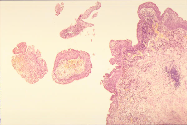
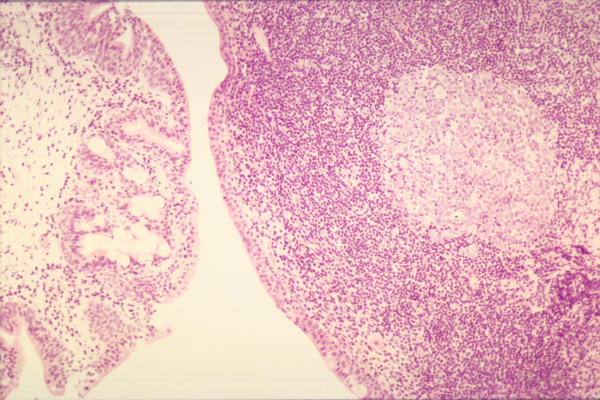
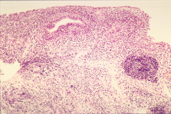
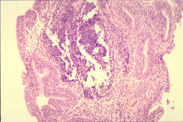
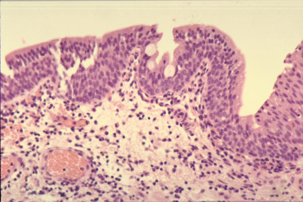
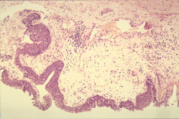
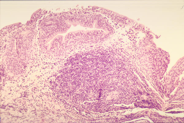
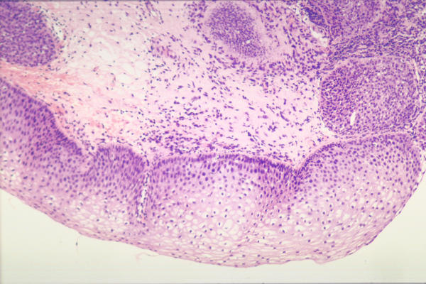
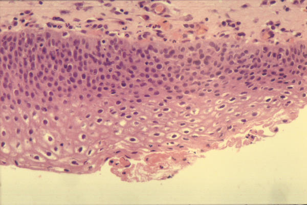
Presentation of the hypothesis: We propose a hypothesis that the type of bladder drainage in SCI patients influences the histological changes in the mucosa of neuropathic bladder. This hypothesis implies that SCI patients with long-term indwelling urinary catheters develop certain histological changes in bladder mucosa, which are seen less frequently in SCI patients, who do not use long-term indwelling catheters. The latter group includes patients, who perform regular intermittent catheterisation and those, who wear a penile sheath and empty their bladders satisfactorily by reflex voiding. We hypothesise that the following histological lesions are seen more frequently in the neuropathic bladder of SCI patients with long-term indwelling catheters.(1) Papillary or polypoid cystitis; (2) widespread cystitis glandularis; (3) moderate to severe, acute and chronic inflammatory changes in bladder mucosa; (4) follicular cystitis; (5) squamous metaplasia; and (6) urothelial dysplasia. As per this hypothesis, it is postulated that the above pathological conditions are seen less often in SCI patients, who achieve complete, low-pressure emptying of the neuropathic bladder by regular intermittent catheterisation, and SCI patients with penile sheath drainage, who empty their bladders satisfactorily by reflex voiding.

Testing the hypothesis: A large prospective study of bladder biopsies in SCI patients practising different methods of bladder drainage is required to validate this hypothesis that the histological changes in bladder mucosa are related to the method of bladder drainage in SCI patients.

Implications of the hypothesis: We propose a hypothesis that the method of bladder drainage in SCI patients influences histological changes in the bladder mucosa. If this hypothesis is validated, methods of bladder drainage such as intermittent catheterisation, which do not require the use of chronic indwelling catheters, should be recommended, in order to minimise adverse histological changes in the mucosa of neuropathic bladder of spinal cord injury patients.


Figures

Figure 1
Figure 1
Photograph of urinary bladder biopsy (H-4268/98): A 54-year male with T-6 paraplegia has been managing the bladder by long-term indwelling urethral catheter. Histopathology shows papillary processes with inflamed fibrovascular cores. The epithelium covering these papillary processes show no cytological atypia. There is severe acute and chronic inflammation with incipient ulceration focally. Large numbers of neutrophils are present within the surface urothelium. The presence of severe inflammation supports the diagnosis of papillary cystitis.
Figure 2
Figure 2
Photograph of urinary bladder biopsy (H-4470/98): A 20-year male sustained C-4 complete tetraplegia two years ago when he fell off football goal post. He has chronic indwelling urethral catheter. Histology shows severe chronic follicular cystitis. There is a large lymphoid follicle with germinal centre, occupying most of the stroma of the biopsy. Elsewhere, there is light scattering of lymphocytes and plasma cells. There is no ulceration of surface urothelium.
Figure 3
Figure 3
Photograph of bladder biopsy (HP-4450/98): A 45-year old male with paraplegia was on penile sheath drainage. He had to bang his stomach to empty the bladder. He had recurrent urinary infections. A staghorn calculus was detected in left kidney during annual review. Cystoscopy revealed mucky, purulent, urine in the bladder. Histology of bladder biopsy revealed moderate to marked chronic inflammation with a minor acute inflammatory component also. The chronic inflammation was predominantly follicular in nature with large lymphoid aggregates including germinal centres, thus giving a diagnosis of follicular cystitis. There was marked cystitis glandularis and focal microcalcification.
Figure 4
Figure 4
Photograph of bladder biopsy (HP00/7187): This 47-year old male sustained C-7 tetraplegia when he was stabbed with a knife in his neck fourteen years ago. He has long-term indwelling urethral catheter. Histology shows cystitis glandularis. There is moderate acute and chronic inflammation without ulceration. The chronic inflammatory infiltrate includes a lymphoid follicle without germinal centre. Polymorphs are present within the surface epithelium
Figure 5
Figure 5
Photograph of bladder biopsy (H-3411/98): A 53-year old male sustained C-5 tetraplegia in a car accident 23 years ago. He has chronic indwelling urethral catheter. Histology shows bladder mucosa covered by predominantly metaplastic epithelium. Only a small amount of normal urothelium is present. The metaplastic epithelium consists of pseudo-stratified columnar cells, with occasional interspersed goblet cells but without cilia. This probably represents a stage in the development of intestinal metaplasia. The stroma contains acute and chronic inflammatory infiltrate.
Figure 6
Figure 6
Photograph of bladder biopsy (H-6004/98): This 18-year old male sustained T-5 paraplegia in a car accident eighteen months ago. He manages his bladder by intermittent catheterisation. He developed bladder stones of unusual shape. The nidus of these stones was hair. Most probably, genital hair was introduced inadvertently into the bladder during self-catheterisation. Histology shows mild chronic inflammation only. There is no acute inflammation or ulceration. The inflammatory infiltrate consists mainly of lymphocytes with occasional plasma cells and mast cells, which is rather patchy and confined to the sub-epithelial zone. Only occasional lymphocytes are present within the urothelium, which is mildly hyperplastic. There is no squamous metaplasia and no evidence of dysplasia. There is also mild oedema and congestion, which may be due to bladder stones.
Figure 7
Figure 7
Photograph of bladder biopsy (H-3640/97): This 49-year old lady developed C-4 tetraplegia three years ago. Since then she has been on long-term indwelling urethral catheter drainage. She developed bladder stone. Histology shows severe acute and chronic inflammation with ulceration. The inflammatory infiltrate includes numerous suburothelial lymphoid follicles. There is focal cystitis glandularis.
Figure 8
Figure 8
Photograph of bladder biopsy (HP00/4013): A 56-year male with T-6 paraplegia has been managing the bladder by long-term indwelling urethral catheter. Histology shows moderate to severe superficial, acute and chronic inflammation without ulceration. Polymorphs extend into the overlying urothelium. A lymphoid follicle with germinal centre is included in the mucosa. Approximately one-third of the surface epithelium shows non-keratinising squamous metaplasia.
Figure 9
Figure 9
Photograph of bladder biopsy (H-5554/99): This 28-year old male developed C-4 complete tetraplegia in a car accident four years ago. He has been on suprapubic catheter drainage for four years. He developed a stone in urinary bladder. Histology shows the bladder mucosa is covered almost entirely by mature, metaplastic squamous epithelium, with only a small amount of transitional epithelium remaining. The squamous epithelium is predominantly non-keratinising. But there are small foci of keratinisation. There is mild dysplasia as well.

References

    1. Yalla SV. Editorial comment on Bladder histological changes associated with chronic indwelling urinary catheter. J Urol. 1999;161:1108–1109. doi: 10.1097/00005392-199904000-00014. - DOI - PubMed
    1. Delnay KM, Stonehill WH, Goldman H, Jukkola AF, Dmochowski RR. Bladder histological changes associated with chronic indwelling urinary catheter. J Urol. 1999;161:1106–1108. doi: 10.1097/00005392-199904000-00013. - DOI - PubMed
    1. Janzen J, Bersch U, Pietsch-Breitfeld B, Pressler H, Michel D, Bultmann B. Urinary bladder biopsies in spinal cord injured patients. Spinal Cord. 2001;39:568–570. doi: 10.1038/sj.sc.3101218. - DOI - PubMed
    1. Hackler RH. Editorial comment on Bladder histological changes associated with chronic indwelling urinary catheter. J Urol. 1999;161:1109. doi: 10.1097/00005392-199904000-00015. - DOI - PubMed
    1. Wall BM, Dmochowski RR, Malecha M, Mangold T, Bobal MA, Cooke CR. Inducible nitric oxide synthase in the bladder of spinal cord injured patients with a chronic indwelling urinary catheter. J Urol. 2001;165:1457–1461. - PubMed

Publication types

MeSH terms