

Review
doi: 10.1186/rr170.
Epub 2002 Apr 4.
Preneoplastic lesions of the lung
Affiliations
- PMID: 11980589
- PMCID: PMC107849
- DOI: 10.1186/rr170
Item in Clipboard
Review
Preneoplastic lesions of the lung
Respir Res.
2002.
Abstract
Lung cancer is the leading cause of cancer deaths worldwide. If we can define and detect preneoplastic lesions, we might have a chance of improving survival. The World Health Organization has defined three preneoplastic lesions of the bronchial epithelium: squamous dysplasia/carcinoma in situ; atypical adenomatous hyperplasia; and diffuse idiopathic pulmonary neuroendocrine cell hyperplasia. These lesions are believed to progress to squamous cell carcinoma, adenocarcinoma and carcinoid tumors, respectively. In this review we summarize the data supporting the preneoplastic nature of these lesions, and delve into some of the genetic changes found in atypical adenomatous hyperplasia and squamous dysplasia/carcinoma in situ.
Figures
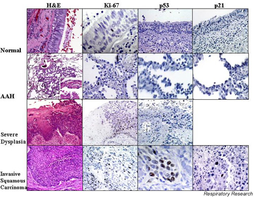
Normal bronchial mucosa, atypical adenomatous hyperplasia (AAH), squamous dysplasia and invasive squamous carcinoma have been stained for various markers. The first column shows hematoxylin and eosin (H&E) tissue staining. The picture of AAH shows low (40×) and high (400×, inset) power images. The H&E staining of squamous dysplasia shows relatively normal mucosa on the left of the image and severe dysplasia on the right. In severe dysplasia, there is considerable cellular pleiomorphism, coarse uneven chromatin, and little cell maturation. Vertical nuclei and mitotic figures are present in the lower two thirds of the mucosa. In invasive carcinoma the cytological aberration is extreme, mitoses occur at all levels of the mucosa, and maturation is absent. The second column shows staining for the proliferation marker, Ki-67, in the various lesions. Ki-67 is essentially negative in normal tissue. The images show increased Ki-67 in AAH, with a further increase in squamous dysplasia and carcinoma. In the third column, p53 is not observed in the normal bronchial mucosa or in AAH, but is increased in both squamous dysplasia and squamous carcinoma. In the fourth column, p21 (p53-inducible cyclin dependent kinase inhibitor) is increased only in squamous carcinoma, but not in AAH or normal bronchial mucosa.
References
-
- Rom WN, Hay JG, Lee TC, Jiang Y, Tchou-Wong KM. Molecular and genetic aspects of lung cancer. Am J Respir Crit Care Med. 2000;161:1355–1367. - PubMed
-
- Patz EF, Jr, Goodman PC, Bepler G. Screening for lung cancer. N Engl J Med. 2000;343:1627–1633. - PubMed
-
- Peck K, Sher YP, Shih JY, Roffler SR, Wu CW, Yang PC. Detection and quantitation of circulating cancer cells in the peripheral blood of lung cancer patients. Cancer Res. 1998;58:2761–2765. - PubMed
-
- Pantel K, Izbicki J, Passlick B, Angstwurm M, Haussinger K, Thetter O, Riethmuller G. Frequency and prognostic significance of isolated tumour cells in bone marrow of patients with non-small-cell lung cancer without overt metastases. Lancet. 1996;347:649–653. - PubMed
-
- Travis CTWD, Corrin B, in collaboration with pathologists from 14 countries . Histological typing of lung and pleural tumours. In: Sobin LH, editor. In World Health Organization International Histological Classification of Tumours. Berlin; New York: Springer-Verlag; 1999.
Publication types
MeSH terms
LinkOut - more resources
Full Text Sources
Medical
