. 2002 May 1;21(9):2122-31.
doi: 10.1093/emboj/21.9.2122.

Export of autotransported proteins proceeds through an oligomeric ring shaped by C-terminal domains

Affiliations

Export of autotransported proteins proceeds through an oligomeric ring shaped by C-terminal domains

Esteban Veiga et al. EMBO J. .

Abstract

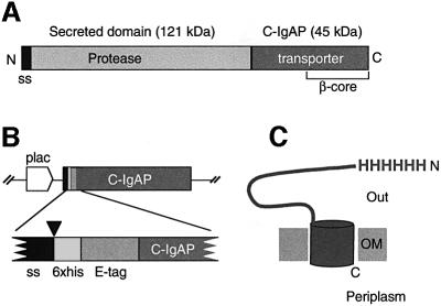
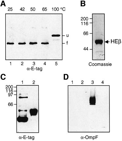
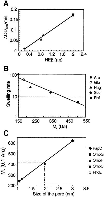
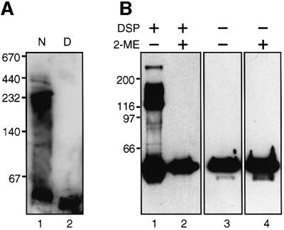
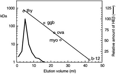
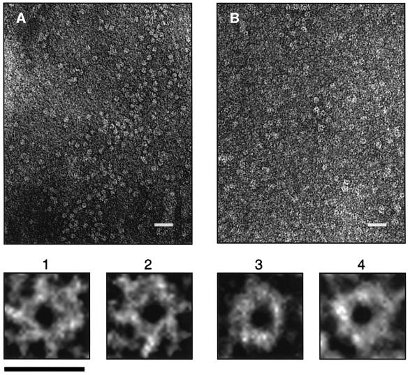
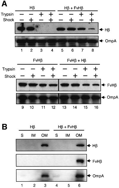
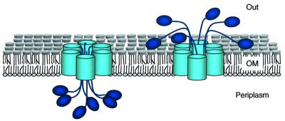
An investigation was made into the oligomerization, the ability to form pores and the secretion-related properties of the 45 kDa C-terminal domain of the IgA protease (C-IgAP) from Neisseria gonorrhoeae. This protease is the best studied example of the autotransporters (ATs), a large family of exoproteins from Gram-negative bacteria that includes numerous virulence factors from human pathogens. These proteins contain an N-terminal passenger domain that em bodies the secreted polypeptide, while the C-domain inserts into the outer membrane (OM) and trans locates the linked N-module into the extracellular medium. Here we report that purified C-IgAP forms an oligomeric complex of approximately 500 kDa with a ring-like structure containing a central cavity of approximately 2 nm diameter that is the conduit for the export of the N-domains. These data overcome the previous model for ATs, which postulated the passage of the N-module through the hydrophilic channel of the beta-barrel of each monomeric C-domain. Our results advocate a secretion mechanism not unlike other bacterial export systems, such as the secretins or fimbrial ushers, which rely on multimeric complexes assembled in the OM.


Figures

None
Fig. 1. Structural organization of the IgA protease and HEβ hybrid. (A) Schematic representation of the IgA protease precursor (∼170 kDa) from N.gonorroheae showing the position of the N-terminal signal sequence (ss), the secreted protease module (∼120 kDa) and the transporter C-domain (∼45 kDa; C-IgAP). The ∼30 kDa β-core region within C-IgAP is also labeled. (B) Organization of the relevant insert of plasmid pHEβ encoding the HEβ hybrid under the control of the IPTG-inducible lac promoter (plac). The heb gene chimera is an in-frame fusion of DNA segments encoding the pelB N-terminal signal sequence (ss), polyhistidine (6×his) and E-tag peptides, and the C-IgAP transporter domain. The site of cleavage of the signal peptidase (just before the His6 peptide) is indicated by an arrowhead. (C) Representation of topology of HEβ in the OM showing the His6 N-passenger domain exposed to the extracellular space (Out) and the C-terminus toward the periplasm. The putative β-barrel of HEβ is depicted as a cylinder embedded in the OM.
None
Fig. 2. Electrophoretic mobility and purification of HEβ. (A) Western blot developed with anti-E-tag mAb–POD of whole-cell protein extracts from IPTG-induced E.coli UT5600 harboring pHEβ. Before loading onto the 10% polyacrylamide gel, the samples were resuspended in denaturing SDS–PAGE sample buffer and heated for 10 min at the indicated temperatures (25, 42, 50, 65 and 100°C). The faster mobility band of HEβ (f) corresponds to the folded conformation of the hybrid. When unfolded, HEβ migrates as a slower mobility band (u). (B) A sample of purified HEβ is shown after Coomassie Blue staining of a 10% SDS–polyacrylamide gel. (C) Western blot developed with anti- E-tag mAb–POD of purified HEβ samples treated at 25 (lane 1) or 100°C (lane 2) for 10 min before loading onto a 10% SDS– polyacrylamide gel. (D) The possible presence of contaminating OmpF porin in the purified HEβ was evaluated by western blot developed with a polyclonal serum against trimeric OmpF. Excess purified HEβ (10 µg, lanes 1 and 2) and a sample of purified OmpF (0.1 µg, lanes 3 and 4) as a control were loaded onto a 10% SDS–polyacrylamide gel after heating at 25 (lanes 1 and 3) or 100°C (lanes 2 and 4) for 10 min. Only the trimeric OmpF control (lane 3) was detected, ruling out the presence of OmpF in the purified HEβ sample.
None
Fig. 3. CD spectrum of HEβ. The CD spectrum of purified HEβ (0.1 mg/ml) was monitored at 22°C in TNZ buffer [20 mM Tris–HCl pH 8.0, 10 mM NaCl, 0.1% (w/v) Zwittergent 3–14]. A minimum of four spectra were accumulated and the contribution of the buffer subtracted. Values of mean residue weight ellipticities (Θ) M.R.W. (degrees × cm2 × dmol–1) are indicated.
None
Fig. 4. Pore-forming activity of HEβ. (A) Swelling rates (ΔOD400/min) of proteoliposomes suspended in isosmotic solutions of arabinose and containing the indicated amount of purified HEβ. (B) Swelling rates of proteoliposomes containing HEβ in solutions of sugars with different Mr. The sugars used were arabinose (Ara; 150 Da), glucose (Glu; 180 Da), N-acetylglucosamine (Nag; 221 Da), sucrose (Suc; 342 Da) and raffinose (Raf; 504 Da). The data are shown relative to the swelling in arabinose and are the averages of at least six independent experiments in which a range of amounts of HEβ (from 0.8 to 2.5 µg) was employed. The swelling rate corresponding to 10% of that in arabinose is indicated with a dashed line. (C) The Mr (0.1 Ara) of HEβ (410 Da) was intersected in a plot representing the Mr (0.1 Ara) of OMPs versus their pore size (PhoE, 240 Da and 1.1 nm; OmpC, 236 Da and 1.1 nm; OmpF, 255 Da and 1.2 nm; OmpG, 400 Da and 2.0 nm; PapC, 620 Da and 3 nm) (Nikaido and Rosenberg, 1983; Cowan et al., 1992; Fajardo et al., 1998; Thanassi et al., 1998b). This plot allows an estimation of 2 nm for the size of HEβ.
None
Fig. 5. Oligomeric forms of C-IgAP in polyacrylamide gels. (A) Western blot probed with anti-E-tag mAb–POD of a native gel containing a 4–15% gradient of polyacrylamide. Purified HEβ was resuspended in native PAGE sample buffer (lane 1, N) or the same buffer containing 1% (w/v) SDS (lane 2, D). The sample in lane 2 was boiled for 10 min before loading. (B) Western blot probed with anti-E-tag mAb–POD of total protein extracts from E.coli UT5600 cells expressing HEβ and incubated in vivo (or not) with DSP cross-linker (lanes 1 and 2). When indicated, the protein extracts were resuspended in SDS–PAGE sample buffer containing 5% (v/v) 2-ME (lanes 2 and 4) in order to reduce a disulfide bridge in the cross-linker. All samples were boiled for 10 min before loading.
None
Fig. 6. Size exclusion chromatography of purified HEβ. The elution profile in a Bio-Gel A column of HEβ and proteins of known Mr as standards [thyroglobulin (thy; 670 000 Da), bovine γ-globulin (ggb; 158 000 Da), chicken ovalbumin (ova; 44 000 Da), equine myoglobin (myo; 17 000 Da) and vitamin B-12 (b-12; 1350 Da)] is shown. Elution of the Mr standards was monitored by UV absorption at 280 nm (open circles), whereas HEβ was detected by western blotting with anti-E-tag mAb–POD.
None
Fig. 7. Electron microscopy of C-IgAP. Electron micrographs (×60 000) of HEβ samples (0.5 mg/ml) stained with (A) 2% ammonium molybdate or (B) 2% uranyl acetate. The white bar corresponds to 35 nm. Images 1 and 2 were magnified from (A), and images 3 and 4 were magnified from (B). The black bar corresponds to 10 nm.
None
Fig. 8. Interference in the translocation of N-domains. (A) The C-IgAP hybrids Hβ and FvHβ were co-expressed (or expressed independently) in E.coli UT5600 cells. These cells were shocked with 10 mM EDTA or incubated with trypsin (1 µg/ml) as indicated (+). The digestion of Hβ and FvHβ was monitored by western blots using anti-His or anti- E-tag mAbs. The amount of OmpA (detected with rabbit anti-OmpA serum) was used as a loading control. (B) The subcellular location of Hβ and FvHβ in E.coli UT5600 cells producing Hβ alone or in combination with FvHβ is shown. The total protein extracts from these E.coli cells were separated into soluble (S), inner membrane (IM) and outer membrane (OM) protein fractions, as described previously (Veiga et al., 1999). OmpA was used as a control of the OM fractions. The proteins were detected by western blot as in (A).
None
Fig. 9. Model for AT secretion. The AT proteins assemble as a ring-shaped oligomeric complex in the bacterial OM. A minimum of six monomers assemble in this complex. The N-terminal passenger domains (P) are depicted as ovals and the C-terminal transporter domain as cylinders anchored in the OM lipid bilayer. The N-domains are translocated from the periplasm (left) to the external medium (right) by passing through the central hydrophilic pore formed by an AT oligomeric complex.

References

    1. Ausubel F.M., Brent,R., Kingston,R.E., Moore,D.D., Seidman,J.G., Smith,J.A. and Struhl,K. (1994) Current Protocols in Molecular Biology. John Wiley & Sons, New York, NY.
    1. Ay J., Keitel,T., Kuttner,G., Wessner,H., Scholz,C., Hahn,M. and Hohne,W. (2000) Crystal structure of a phage library-derived single-chain Fv fragment complexed with turkey egg-white lysozyme at 2.0 Å resolution. J. Mol. Biol., 301, 239–246. - PubMed
    1. Benz I. and Schmidt,M.A. (1992) AIDA-I, the adhesin involved in diffuse adherence of diarrhoeagenic Escherichia coli strain 2787 (O126:H27), is synthesized via a precursor molecule. Mol. Microbiol., 6, 1539–1546. - PubMed
    1. Bernstein H.D. (2000) The biogenesis and assembly of bacterial membrane proteins. Curr. Opin. Microbiol., 3, 203–209. - PubMed
    1. Bitter W., Koster,M., Latijnhouwers,M., de Cock,H. and Tommassen,J. (1998) Formation of oligomeric rings by XcpQ and PilQ, which are involved in protein transport across the outer membrane of Pseudomonas aeruginosa. Mol. Microbiol., 27, 209–219. - PubMed

Publication types

MeSH terms