. 2002 May 1;21(9):2231-41.
doi: 10.1093/emboj/21.9.2231.

WSTF-ISWI chromatin remodeling complex targets heterochromatic replication foci

Affiliations

WSTF-ISWI chromatin remodeling complex targets heterochromatic replication foci

Ludmila Bozhenok et al. EMBO J. .

Erratum in

  • EMBO J 2002 Jun 17;21(12):3212

Abstract

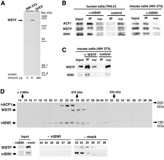
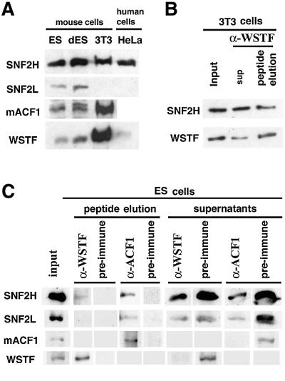
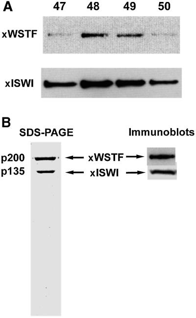
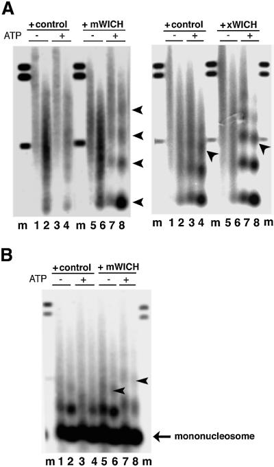
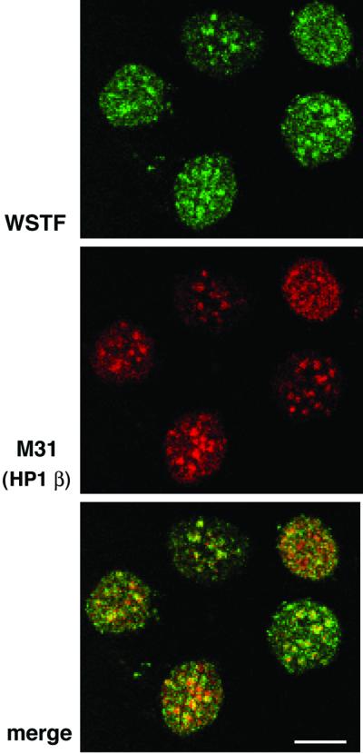
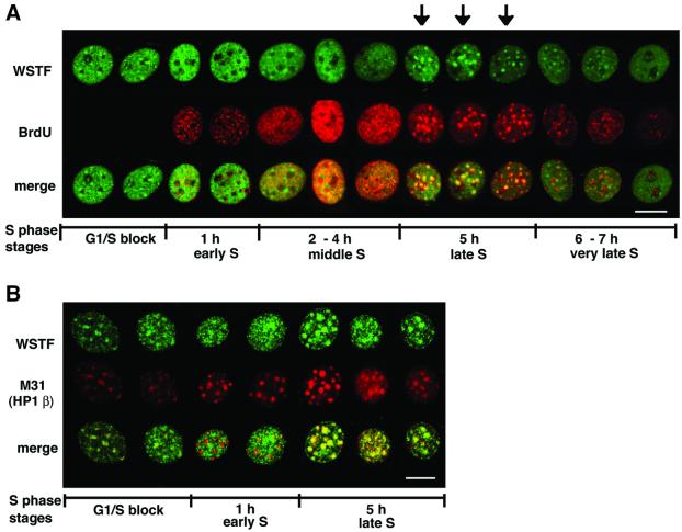
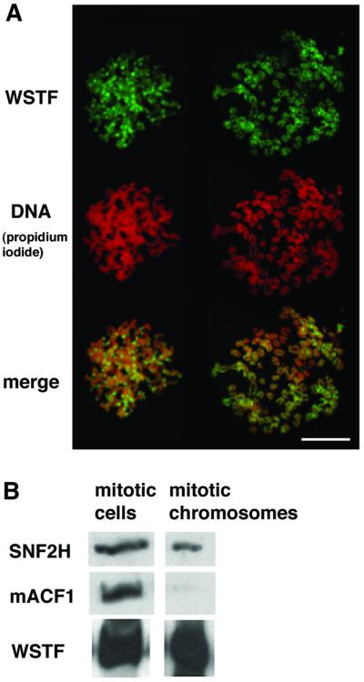
The Williams Syndrome Transcription Factor (WSTF), the product of the WBSCR9 gene, is invariably deleted in the haploinsufficiency Williams-Beuren Syndrome. Along with the nucleosome-dependent ATPase ISWI, WSTF forms a novel chromatin remodeling complex, WICH (WSTF-ISWI chromatin remodeling complex), which is conserved in vertebrates. The WICH complex was purified to homogeneity from Xenopus egg extract and was found to contain only WSTF and ISWI. In mouse cells, WSTF interacts with the SNF2H isoform of ISWI. WSTF accumulates in pericentric heterochromatin coincident with the replication of these structures, suggesting a role for WSTF in the replication of heterochromatin. Such a role is supported by the in vitro activity of both the mouse and frog WICH complexes: they are involved in the assembly of regular spaced nucleosomal arrays. In contrast to the related ISWI-interacting protein ACF1/WCRF180, WSTF binds stably to mitotic chromosomes. As dysfunction of other chromatin remodeling factors often has severe effects on development, haploinsufficiency of WSTF may explain some of the phenotypes associated with this disease.


Figures

None
Fig. 1. WSTF is conserved in vertebrates. The domain structure of WSTF is shown above. Bars below show the matching positions of corresponding ESTs from X.laevis (1–4), zebrafish (D.rerio, 5–7) and S.tropicalis (8–9). Accession numbers: 1, BI447904; 2, BG264264; 3, BI447594; 4, BG345707; 5, AI794397; 6, AI436874; 7, AW466480; 8, BG514920; 9, BG515236.
None
Fig. 2. WSTF and ISWI form a complex in mouse and human cells. (A) Western blot analysis of NIH 3T3 nuclear extract and Drosophila embryo extract using affinity-purified anti-WSTF antibody. (B) Co-immunoprecipitation of WSTF with affinity-purified anti-ISWI antibodies in HeLa cell nuclear extracts (left panel) and NIH 3T3 cell nuclear extracts (right panel). Input (12%); IP, immunoprecipitate (100%); sup, supernatant of immunoprecipitate. (C) Co-immunoprecipitation of ISWI with affinity-purified anti-WSTF antibodies. Input (4%); IP, immunoprecipitate (100%). Control immunoprecipitations in (B) and (C) were with the same amount of purified rabbit IgG. (D) Fractionation of WSTF, hACF1(WCRF180) and ISWI from crude HeLa nuclear extract by Superose-6 gel filtration chromatography. Upper panel: overlay of three separate western blots against WSTF, hACF1 (WCRF180) and hISWI of the fractions. Lower panel: fractionation of WSTF in HeLa nuclear extract immunodepleted with antibodies against hISWI (mock depletion was with pre-immune serum). Size standards were thyroglobulin (670 kDa) and catalase (232 kDa).
None
Fig. 3. WSTF interacts with the SNF2H isoform of ISWI. (A) Western blot of identical amounts of nuclear extract proteins from mouse ES cells, ES cells driven to differentiation (dES), NIH 3T3 and HeLa cells. (B) Western blot of affinity-purified mouse WSTF–ISWI complex: input, 10%; peptide eluate, 100%. (C) Immunoprecipitation of WSTF– and ACF1–ISWI complexes from ES cell nuclear extract. The complexes were eluted from the antibodies with the antigenic peptides and analyzed by western blots: input, 16%; peptide eluate, 100%.
None
Fig. 4. WSTF forms a complex with ISWI in Xenopus egg extracts. (A) Western blot of co-purification of Xenopus WSTF (anti-human WSTF antibodies) and Xenopus ISWI (anti-Xenopus ISWI antibodies) over MonoS chromatography during ISWI-B purification. This is the penultimate step in the purification scheme. (B) SDS–PAGE of the final purified complex showing the two polypeptides. Immunoblot shows the antibody reactivity with the respective antisera.
None
Fig. 5. The WSTF–ISWI complex is a chromatin remodeling factor, WICH. (A) Left panel: the mouse WSTF–ISWI complex reconfigures irregular chromatin into a regular nucleosomal array. Eluates from the pre-immune serum (+control) or anti-WSTF antiserum (+WICH) were added to sarcosyl-stripped chromatin, assembled in Drosophila embryo extracts without ATP. Addition of 1 mM ATP was as indicated. Micrococcal nuclease digestion was for 30 s (lanes 1, 3, 5 and 7) or 60 s (lanes 2, 4, 6 and 8). Lanes labeled ‘m’ contain size marker DNA fragments: 0.49, 1.1 and 1.2 kbp. Arrows indicate mono-, di-, tri- and tetranucleosome DNA fragments (from the bottom upwards). Right panel: same experiment as in the left panel, but with the purified Xenopus WICH (300 ng MonoS fraction, purification buffer as control). Arrows indicate the position of the trinucleosome DNA fragment. (B) WICH mobilizes nucleosomes. Nucleosomes were assembled with polyglutamic acid as carrier on plasmid DNA. This chromatin was incubated with mock eluate (+control) or the immunopurified WSTF complex (+mWICH). Addition of 1 mM ATP was as indicated above the panels. Micrococcal nuclease digestion was for 30 s (lanes 1, 3, 5 and 7) or 60 s (2, 4, 6 and 8). Lanes labeled ‘m’ contain size marker DNA fragments: 0.49, 1.1 and 1.2 kbp. Arrows indicate a change of internucleosomal repeat length.
None
Fig. 6. WSTF co-localizes with M31 (mouse HP1β), a marker protein for mouse pericentromeric heterochromatin. NIH 3T3 cells were fixed and stained with affinity-purified anti-WSTF antibodies (top, FITC, green) and rat monoclonal antibodies against M31 (middle, Texas Red). Lower panel: merged image (yellow). Bar = 10 µm.
None
Fig. 7. WSTF is targeted to pericentromeric heterochromatin during its replication. (A) NIH 3T3 cells were synchronized at the G1/S border, released into S phase, fixed at the indicated times after release and stained for WSTF (top, FITC, green) and BrdU (middle, Texas Red) incorporation. The merged image (yellow) is shown at the bottom. Arrows indicate cells with WSTF foci at late S phase. (B) WSTF (top, green) and M31 (HP1β, middle, red) staining through S phase. Bottom panel: merged image (yellow). Bar = 10 µm.
None
Fig. 8. WSTF binds mitotic chromosomes. (A) Anti-WSTF staining (FITC, green) of spread mitotic chromosomes from NIH 3T3 cells, and propidium iodide staining for DNA (red, middle). Bottom: merged image (yellow). Bar = 10 µm. (B) Western blot analysis of whole mitotic NIH 3T3 cells and purified mitotic chromosomes. Each sample contains the same amount of chromatin.

References

    1. Aihara T., Miyoshi,Y., Koyama,K., Suzuki,M., Takahashi,E., Monden,M. and Nakamura,Y. (1998) Cloning and mapping of SMARCA5 encoding hSNF2H, a novel human homologue of Drosophila ISWI. Cytogenet. Cell Genet., 81, 191–193. - PubMed
    1. Alexiadis V., Varga-Weisz,P.D., Becker,P.B. and Gruss,C. (1998) In vitro chromatin remodeling by chromatin accessibility complex (CHRAC) at the SV40 origin of DNA replication. EMBO J., 17, 3428–3438. - PMC - PubMed
    1. Altschul S.F., Madden,T.L., Schaffer,A.A., Zhang,J., Zhang,Z., Miller,W. and Lipman,D.J. (1997) Gapped BLAST and PSI-BLAST: a new generation of protein database search programs. Nucleic Acids Res., 25, 3389–3402. - PMC - PubMed
    1. Amir R.E., Van den Veyver,I.B., Wan,M., Tran,C.Q., Francke,U. and Zoghbi,H.Y. (1999) Rett syndrome is caused by mutations in X-linked MECP2, encoding methyl-CpG-binding protein 2. Nature Genet., 23, 185–188. - PubMed
    1. Bochar D.A., Savard,J., Wang,W., Lafleur,D.W., Moore,P., Cote,J. and Shiekhattar,R. (2000) A family of chromatin remodeling factors related to Williams syndrome transcription factor. Proc. Natl Acad. Sci. USA, 97, 1038–1043. - PMC - PubMed

Publication types