

Comparison of an immortalized human corneal epithelial cell line with Vero cells in the isolation of Herpes simplex virus-1 for the laboratory diagnosis of Herpes simplex keratitis
- PMID: 11983023
- PMCID: PMC113264
- DOI: 10.1186/1471-2415-2-3
Comparison of an immortalized human corneal epithelial cell line with Vero cells in the isolation of Herpes simplex virus-1 for the laboratory diagnosis of Herpes simplex keratitis
Abstract
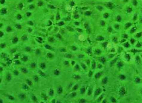
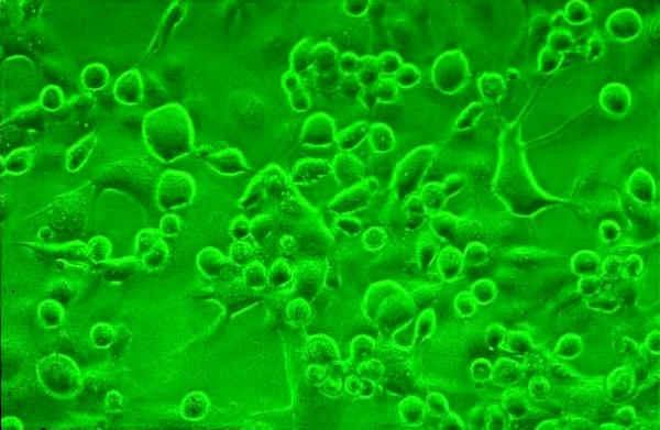
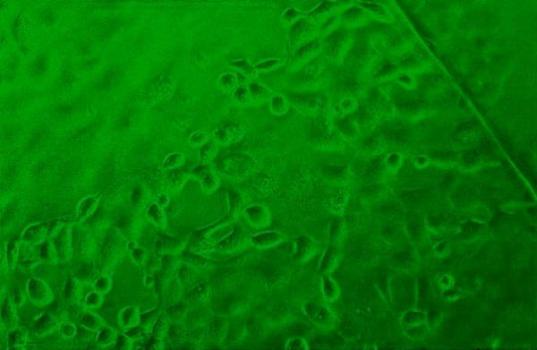
Background: Herpes simplex keratitis (HSK) is a sight threatening ocular infection often requiring a specific and prompt laboratory diagnosis. Isolation of Herpes simplex virus (HSV-1) in culture provides the most reliable and specific method and is considered as the "Gold Standard" in the laboratory diagnosis of HSK in spite of its low sensitivity. Using "cell lines of corneal origin" for virus isolation may be beneficial under such circumstances, since these cells have been shown to be excellent substrates for the growth of HSV-1 isolated from the cornea. We report a comparative study of a novel human corneal epithelial cell line (HCE) and the Vero cell line in the isolation of HSV-1 from corneal scrapings employing a shell vial assay.
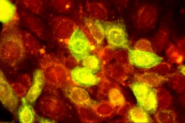
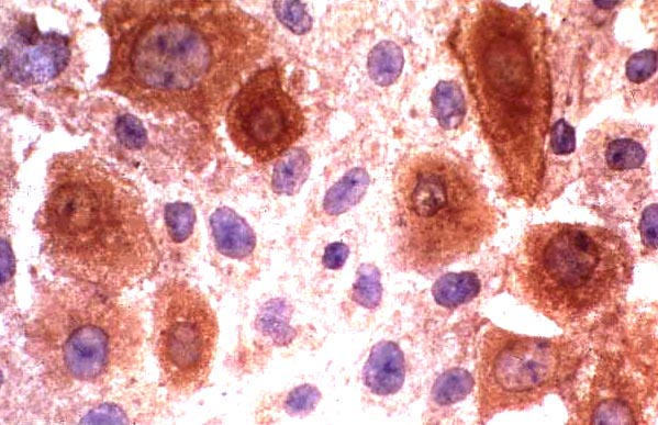
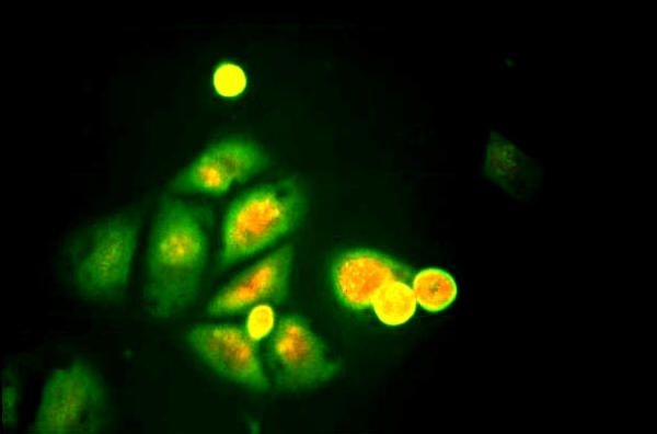
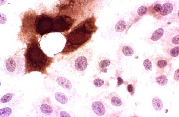
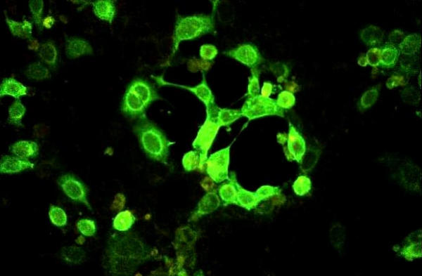
Methods: Corneal scrapings were obtained from 17 patients with a clinical diagnosis of HSK. All the cases were confirmed by virological investigations (PCR and viral antigen detection positive, n = 15, PCR positive, n = 1, Viral antigen positive, n = 1). Scrapings obtained from 10 patients with infectious keratitis of non-viral origin were included as controls. All the scrapings were simultaneously inoculated into shell vials of HCE and Vero cells. Cultures were terminated at 24 h post-infection. Isolation of HSV-1 was confirmed using an indirect immunofluorescence/ immunoperoxidase assay.
Results: Virus could be isolated using both or either of the cell lines in 10/17 (58.82%) patients with HSK. HSV-1 was isolated from 10/17 (58.82%) and 4/17(23.52%) specimens in HCE and Vero cells, respectively (P = 0.036). None of the controls yielded HSV-1. While all the 10 (100%) strains were isolated in HCE, Vero yielded only 4/10 (40%) strains in the shell vial culture (P = 0.014).
Conclusions: HCE showed a statistically significant difference in the virus isolation rate with respect to Vero cells. HCE may be an excellent alternative cell line for the isolation of HSV-1, especially from corneal scrapings, for the laboratory diagnosis of HSK.
Figures
References
-
- Yamamoto SY, Shimomura Y, Kinoshita S, Nishida K, yamamoto R, Tano Y. Detection of herpes simplex virus DNA in human tear film by polymerase chain reaction. Am J Ophthalmol. 1994;117:160–163. - PubMed
-
- Kowalski RP, Gordon YJ. Evaluation of immunologic tests for the detection of ocular herpes simplex virus. Ophthalmology. 1989;96:1583–1586. - PubMed
Publication types
MeSH terms
Substances
LinkOut - more resources
Full Text Sources
