. 2002 May 14;99(10):6901-6.
doi: 10.1073/pnas.092148299. Epub 2002 Apr 30.

Global changes in gene expression by human polymorphonuclear leukocytes during receptor-mediated phagocytosis: cell fate is regulated at the level of gene expression

Affiliations

Global changes in gene expression by human polymorphonuclear leukocytes during receptor-mediated phagocytosis: cell fate is regulated at the level of gene expression

Scott D Kobayashi et al. Proc Natl Acad Sci U S A. .

Abstract

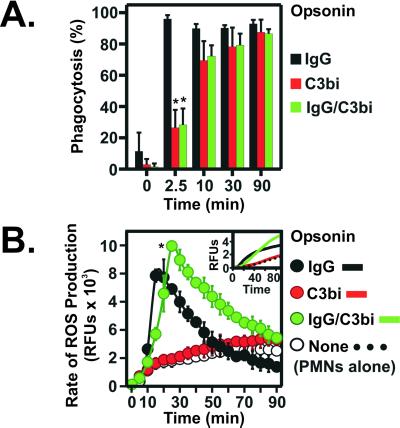
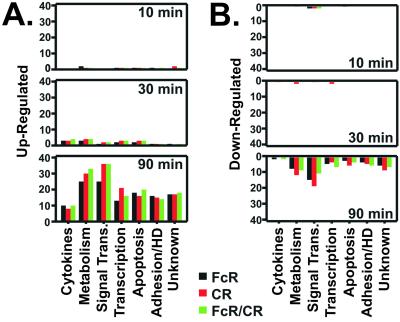
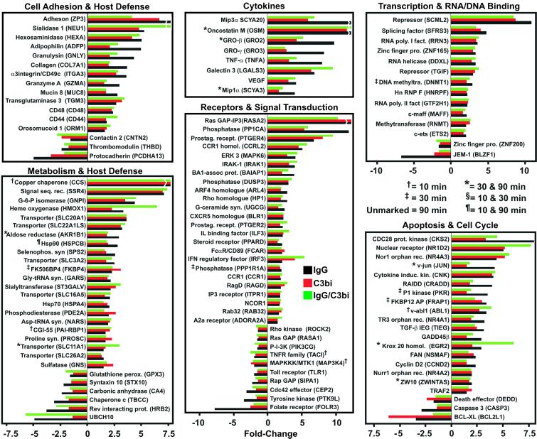
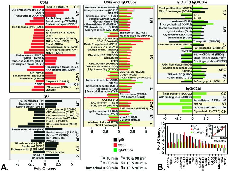
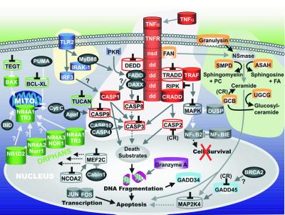
Phagocytes are a critical component of the innate immune response in humans and eliminate invading microorganisms through a process known as phagocytosis. Two distinct receptor-linked phagocytic pathways, one with Ab receptors (FcRs; FcR, Fc receptor) and the other complement receptors (CRs), mediate binding and ingestion of pathogens by human polymorphonuclear leukocytes (PMNs). Although progress has been made toward defining complex signal transduction processes that underlie phagocytosis in each pathway, very little is known about gene regulation during or after phagocytosis. Therefore, we used human oligonucleotide microarrays to identify changes in expression of 12,561 genes accompanying FcR- and CR-mediated phagocytosis. Eighty-four percent of 279 differentially expressed genes were induced or repressed 90 min after ingestion of Ab- and/or complement-opsonized particles. Unexpectedly, more than 30 of these genes encoded proteins involved in at least three distinct apoptotic pathways. Ninety-four differentially expressed cell fate-related genes were identified between 180 and 360 min after phagocytosis and most were induced or repressed by PMNs activated through both receptors simultaneously. By using flow cytometry, we found that FcR- and CR-mediated phagocytosis each promoted programmed cell death in human PMNs; however, phagocytosis mediated by the combination of FcRs and CRs induced apoptosis earlier than that by either receptor alone. Our results reveal distinct patterns of receptor-mediated gene expression that define complex inducible apoptotic pathways in activated PMNs. Most significantly, we discovered that programmed cell death is regulated at the level of gene expression. Thus, we hypothesize that gene regulation in PMNs facilitates resolution of inflammatory responses.


Figures

Figure 1
Figure 1
FcR- and CR-mediated phagocytosis in human PMNs. (A) PMNs were stimulated with IgG LB (IgG), C3bi LB (C3Bi), or IgG/C3bi LB (IgG/C3bi), and phagocytosis was quantitated as described in Materials and Methods. Results are the mean ± SD of 3–5 separate experiments. *, P < 0.01 vs. IgG LB. (B) ROS production during phagocytosis. Data are expressed as rate of ROS production over time and are the mean ± SD of 3–6 experiments. Inset illustrates representative plots of FcR-, CR-, and FcR/CR-stimulated ROS production. *, P < 0.01 vs. PMNs stimulated with IgG and C3bi LB.
Figure 2
Figure 2
Differential gene expression during phagocytosis. (A and B) Human PMNs were stimulated with IgG LB (FcR), C3bi LB (CR), or IgG/C3bi LB (FcR/CR) for the indicated times, and gene expression was measured with Affymetrix human oligonucleotide microarrays. Data were analyzed with GENESPRING software version 4.0.4 (Silicon Genetics, Redwood City, CA). Genes induced (A) or repressed (B) after FcR- (black bars), CR- (red bars), or FcR/CR-mediated phagocytosis (green bars) were categorized by function and enumerated. HD, host defense.
Figure 3
Figure 3
Differentially expressed genes shared between FcR- and CR-mediated phagocytosis. Genes differentially expressed during and after FcR- (black bars), CR- (red bars), or FcR/CR-mediated phagocytosis (green bars) were categorized by function. Breaks in the bars indicate genes whose differential expression level is off-scale. Results are presented as the mean fold-induction or repression of genes from three experiments.
Figure 4
Figure 4
Receptor-specific induction or repression of genes during phagocytosis. (A) Differentially expressed genes identified in both C3bi and IgG/C3bi (CR-specific) or IgG and IgG/C3bi (FcR-specific) stimulated PMNs are labeled as C3bi and IgG/C3bi and IgG and IgG/C3bi, respectively. CC, cytokines, chemokines, and growth factors; MT, metabolism and trafficking; ST, receptors and signal transduction; T, transcription and RNA/DNA binding; APO, apoptosis and cell cycle; CH, cell adhesion and host defense; ?, unknown function. (B) Taqman verification of microarray results. Genes (n = 19) identified as differentially transcribed by Affymetrix microarrays were selected for confirmation by Taqman real-time PCR. Inset illustrates the correlation (r = 0.81) between real-time PCR and microarray analyses.
Figure 5
Figure 5
Phagocytosis-induced apoptosis in PMNs. (A) Induction or repression of cell fate-related genes in PMNs after phagocytosis was determined 180 (†) and 360 (‡) min after phagocytosis as indicated. Genes differentially expressed at least 2-fold are shaded in gray. *, P < 0.05 for stimulated (FcR, CR, and FcR/CR) vs. unstimulated PMNs. (B) Flow cytometric analysis of early apoptosis during phagocytosis. PMNs with exposed phosphatidylserine (early apoptosis) bound Annexin V-FITC (lower right quadrants); upper quadrants, propidium iodide staining. Percent staining is indicated within the quadrant, and data are representative of six separate experiments. (C) After phagocytosis, chromatin condensation in apoptotic PMNs was determined by flow cytometry. Percent of apoptotic nuclei is indicated within the histogram panels. (D) After phagocytosis, late apoptosis (DNA fragmentation) in PMNs was detected with a modified terminal deoxynucleotidyltransferase dUTP nick end-labeling assay. Percent DNA fragmentation is indicated within each histogram panel. Results are representative of three separate experiments. (E) Quantitation of six apoptosis (chromatin condensation) experiments. *, P < 0.01 vs. FcR-, CR-phagocytosis, and unstimulated PMNs (CTL).
Figure 6
Figure 6
Model of phagocytosis-induced cell fate in human PMNs. Apoptosis-related genes identified in this study were categorized into signaling pathways involved in cell fate; tumor necrosis factor-receptor (red), ceramide synthesis (light orange), Toll-like receptor signaling (dark blue), mitochondrial (green), or nuclear (gray). Genes up-regulated are illustrated as solid cylinders, genes down-regulated are illustrated as open cylinders, and genes expressed but neither induced nor repressed are depicted by dark gray spheres. Genes down-regulated also contain an inverted black “T” and in some instances repression or induction by a specific receptor is indicated (in parentheses). Solid lines/arrows indicate positive effector functions; dotted lines/arrows indicate the absence of effector functions; gray lines ending with an inverted T indicate block of function. The model is based on gene and protein functions reported in the literature, but has been modified to fit our data set.

References

    1. Nauseef W M, Clark R A. In: Basic Principles in the Diagnosis and Management of Infectious Diseases. Mandell G L, Bennett J E, Dolin R, editors. New York: Churchill Livingstone; 2000. pp. 89–112.
    1. Daeron M. Annu Rev Immunol. 1997;15:203–234. - PubMed
    1. Sengelov H. Crit Rev Immunol. 1995;15:107–131. - PubMed
    1. Greenberg S, Chang P, Silverstein S C. J Exp Med. 1993;177:529–534. - PMC - PubMed
    1. Cox D, Tseng C C, Bjekic G, Greenberg S. J Biol Chem. 1999;274:1240–1247. - PubMed