. 2002 May 14;99(10):6973-8.
doi: 10.1073/pnas.102657399. Epub 2002 Apr 30.

beta-Catenin stabilization dysregulates mesenchymal cell proliferation, motility, and invasiveness and causes aggressive fibromatosis and hyperplastic cutaneous wounds

Affiliations

beta-Catenin stabilization dysregulates mesenchymal cell proliferation, motility, and invasiveness and causes aggressive fibromatosis and hyperplastic cutaneous wounds

Sophia S Cheon et al. Proc Natl Acad Sci U S A. .

Abstract

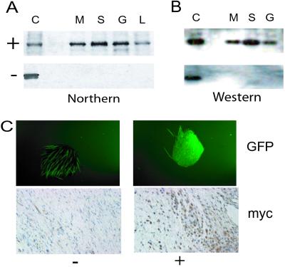
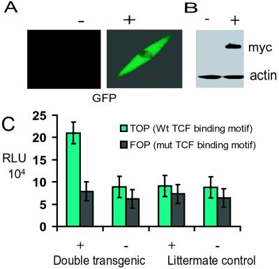
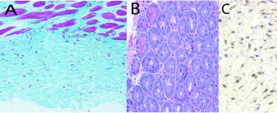
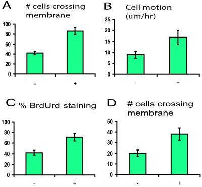
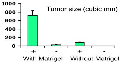
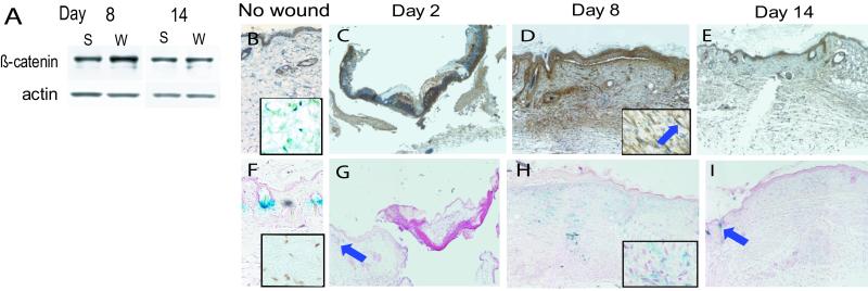
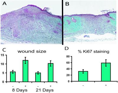
Fibroproliferative processes are a group of disorders in which there is excessive proliferation of spindle (mesenchymal fibroblast-like) cells. They range from hypertrophic scars to neoplasms such as aggressive fibromatosis. Cells from these disorders share cytologic similarity with fibroblasts present during the proliferative phase of wound healing, suggesting that they represent a prolonged wounding response. A critical role for beta-catenin in mesenchymal cells in fibroproliferative processes is suggested by its high rate of somatic mutation in aggressive fibromatosis. Using a Tcf-reporter mouse we found that beta-catenin protein level and Tcf-transcriptional activity are elevated in fibroblasts during the proliferative phase of healing. We generated a transgenic mouse in which stabilized beta-catenin is expressed in mesenchymal cells under control of a tetracycline-regulated promoter. Fibroblasts from the transgenic mice exhibited increased proliferation, motility, and invasiveness when expressing stabilized beta-catenin and induced tumors after induction of the transgene when grafted into nude mice. Mice developed aggressive fibromatoses and hyperplastic gastrointestinal polyps after 3 months of transgene induction and healed with hyperplastic cutaneous wounds compared with control mice, which demonstrates an important function for beta-catenin in mesenchymal cells and shows a central role for beta-catenin in wound healing and fibroproliferative disorders.


Figures

Figure 1
Figure 1
Regulation of the transgene by doxycycline in double-transgenic mice. (A) Northern analysis using a transgene-specific probe. +, mice treated with doxycycline; −, mice not treated; C, positive control; M, muscle; S, skin; G, gastrointestinal tract; L, liver. (B) Western analysis using an anti-Myc-epitope antibody on immunoprecipitates performed with an anti-β-catenin antibody on protein extracts. (C) Regulation of transgene expression as demonstrated by green fluorescent protein production in a full-thickness punch sample from a mouse ear (Upper, autofluorescence of hair is noted in the untreated sample) and by immunohistochemistry using an anti-Myc-epitope antibody in dermal tissue (Lower). Strict regulation of expression of the transgene by doxycycline administration is demonstrated.
Figure 2
Figure 2
Tcf-dependent transcriptional activation in primary fibroblast cell cultures. +, treated with doxycycline; −, not treated. The regulation of transgene expression in primary cell cultures is shown. A shows green fluorescent protein production, and B shows Western analysis using an anti-Myc-epitope antibody with actin as a loading control. (C) Tcf-transcriptional activation (pTOPFLASH) is increased significantly with induction on the transgene, compared with activation of a control reporter construct (pFOPFLASH) or control cell cultures. RLU, relative luciferase units; Wt, wild type; mut, mutant.
Figure 3
Figure 3
Histology of tumors formed in the mice. (A) An aggressive fibromatosis with spindle cells infiltrating into the muscle tissue. (B) A hyperplastic gastrointestinal polyp. (C) Immunohistochemistry using an anti-Myc-epitope antibody in an aggressive fibromatosis, illustrating expression of the transgene in the tumor.
Figure 4
Figure 4
Result of transgene stabilization on behavior of primary fibroblast cultures. +, treated with doxycycline; −, not treated. (A) Motility as measured by the number of cells per high-powered field crossing the membrane using a modified Boyden chamber. (B) Cell motion as measured by using time-lapse photomicroscopy. (C) Proliferation as measured by using the percentage of cells incorporating BrdUrd. (D) Cell invasiveness as measured by the number of cells per high-power field crossing Matrigel. In all cases, activation of the transgene resulted in a significant increase.
Figure 5
Figure 5
Size of tumors formed with cells grafted into nude mice. +, treated with doxycycline; −, not treated. A significantly increased size is noted with induction of the transgene, with or without the use of Matrigel.
Figure 6
Figure 6
β-Catenin-mediated tcf-dependent transcription is activated in the proliferative phase of wound healing. (A) Western analysis from wounds 8 and 14 days after wounding, showing an increase in β-catenin protein in the wound at day 8 (actin staining as a loading control). W, wound tissue; S, normal tissue. (BE) β-Catenin immunohistochemistry, showing increased staining in the mesenchymal tissues (magnified view in box with arrow pointing to a fibroblast expressing β-catenin) at day 8 (D). (FI) Tcf-activation in the reporter mouse. Blue staining indicates tcf activation. The arrows point to hair follicles, where tcf is known to be activated, as a positive control. The 8-day sample (H) shows tcf activation in the fibroblasts, better illustrated in the magnified view in the box. β-Catenin-mediated tcf-dependent activation in hair follicles, illustrated in the unwounded skin shown in F and by the arrow in G, may result in underestimation of the actual difference in β-catenin protein level when comparing with unwounded skin, because hair follicles are not present in wound tissue. The insets in B and F show a lack of β-catenin-mediated tcf-dependent activation in dermal fibroblasts in unwounded skin.
Figure 7
Figure 7
Wounds are larger in animals expressing the stabilized β-catenin. A histologic section taken through the middle of the wound 8 days after wounding. A larger size to the mesenchymal component of the wound in the mouse treated with doxycycline (A) is illustrated compared with a transgenic mouse not treated with doxycycline (B). In this particular sample, a thicker epithelial component is present also; however, there was not a significant difference in size of the epithelial component when taking all the samples into account. (C) Graphic representation of the difference in wound area between treated (+) and untreated (−) animals at 8 and 21 days after wounding. (D) Difference in Ki-67 staining (by percentage of cells) between wounds in mice receiving doxycycline (+) and those not receiving doxycycline (−) at 8 days after wounding.

References

    1. Polakis P. Genes Dev. 2000;14:1837–1851. - PubMed
    1. Taipale J, Beachy PA. Nature (London) 2001;411:349–354. - PubMed
    1. Korswagen HC, Clevers HC. Cold Spring Harbor Symp Quant Biol. 1999;64:141–147. - PubMed
    1. Lattes R. In: Tumors of the Soft Tissues. Lattes R, editor. Washington, DC: Armed Forces Institute of Pathology; 1980. pp. 1–30.
    1. Epstein F H. N Engl J Med. 1999;341:738–746. - PubMed

Publication types

MeSH terms

LinkOut - more resources