. 2002 Apr 30;99(9):5925-30.
doi: 10.1073/pnas.082120199.

A chloroplast-resident DNA methyltransferase is responsible for hypermethylation of chloroplast genes in Chlamydomonas maternal gametes

Affiliations

A chloroplast-resident DNA methyltransferase is responsible for hypermethylation of chloroplast genes in Chlamydomonas maternal gametes

Rie Nishiyama et al. Proc Natl Acad Sci U S A. .

Abstract

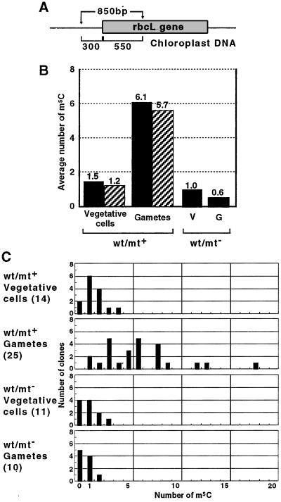
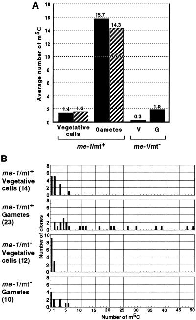
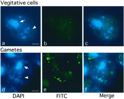
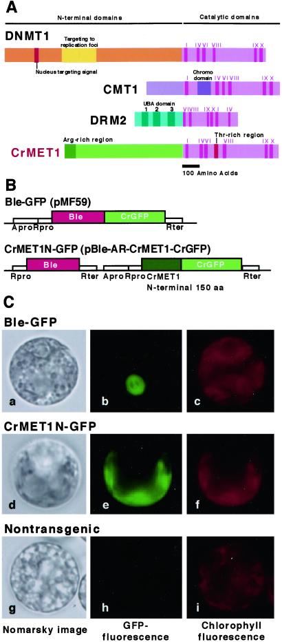
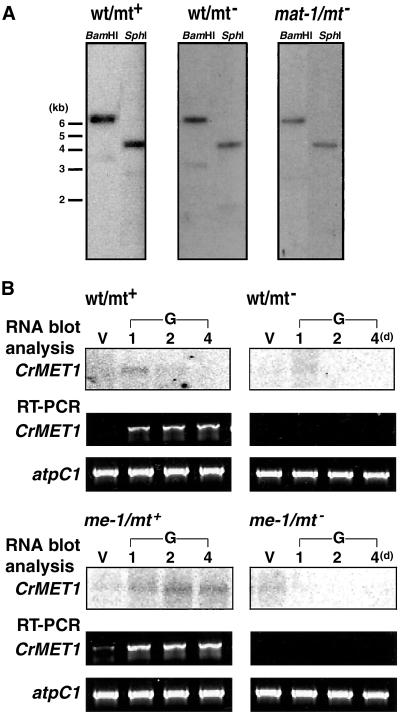
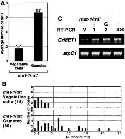
Chloroplast DNA of the green alga Chlamydomonas reinhardtii is maternally inherited. Methylation mapping directly revealed that, before mating, chloroplast DNA of maternal (mating type plus; mt(+)) gametes is heavily methylated whereas that of paternal (mating type minus; mt(-)) gametes is not. Indirect immunofluorescence analyses with anti-5-methylcytosine mAbs visually showed methylation to occur exclusively in chloroplast DNA of mt(+) gametes, and not in mt(-) gametes or nuclear DNA of either mt. To clarify the relationship between methylation and maternal inheritance of chloroplast DNA, we have isolated and characterized a cDNA encoding a DNA methyltransferase. The deduced protein, CrMET1, consists of 1,344 aa and contains a conserved catalytic domain at the C terminal and a nonconserved N-terminal region. The predicted N-terminal region has an arginine-rich domain, suggesting CrMET1 is transferred to chloroplasts. This finding could be directly shown by green fluorescent protein epifluorescence microscopy analyses. CrMET1 transcripts were found to be absent in both mt(+) and mt(-) vegetative cells. Upon gametogenesis, however, transcript levels clearly increased in mt(+) but not mt(-) cells. These experiments suggest that the CrMET1 protein is located in chloroplasts and that it specifically methylates cytosine residues of chloroplast DNA in mt(+) gametes. This conclusion was further strengthened by the observation that, during gametogenesis, CrMET1 is expressed in a mt(-) mutant, mat-1, whose chloroplast DNA is heavily methylated in gametes and paternally inherited. The results provide evidence that cytosine methylation plays a critical role in maternal inheritance of chloroplast genes in C. reinhardtii.


Figures

Figure 1
Figure 1
Methylation mapping of the rbcL gene in wild-type (wt) cells. (A) Analyzed region of the rbcL locus in chloroplast DNA. The 850 bp contains 300 bp and 550 bp of promoter and coding regions, respectively. The total number of cytosine residues is 149 in the A strand (sense strand) and 148 in the B strand (antisense strand). (B) Average numbers of m5C. After 30 cycles of PCR (94°C 15 sec, 50°C 15 sec, 72°C 1 min) using 10 ng of the bisulfite-modified DNA, the products were digested by SalI and SacI, gel-purified, and cloned into pBluescript KS−. Individual clones were sequenced and sites of methylated cytosines were identified. The numbers of sequenced clones were 14 and 11 for mt+ and mt vegetative cells, respectively, and 25 and 10 for mt+ and mt gametes, respectively. Average numbers of m5C in A and B strands are indicated by solid and hatched bars, respectively. (C) Numbers of m5C in individual clones. The value for a given clone is indicated on the horizontal scale, and the number of clones containing the indicated m5C number is shown on the vertical scale. The sequence specificity of the m5C is not shown, but 36.8% of the total m5C appeared in CpG, 27.6% in CpNpG, and 35.5% in other sequences, apparently at random.
Figure 2
Figure 2
Methylation mapping of the rbcL gene in me-1 mutant cells. (A) Average numbers of m5C. Experiments were performed as described in the legend for Fig. 1. The numbers of sequenced clones were 14 and 12 for mt+ and mt vegetative cells, respectively, and 23 and 10 for mt+ and mt gametes, respectively. Average numbers of m5C in A and B strands are indicated by solid and hatched bars, respectively. (B) Numbers of m5C in individual clones. See the legend for Fig. 1. Of the total m5C, 38.4% appeared in CpG, 14.1% in CpNpG, and 47.5% in other sequences.
Figure 3
Figure 3
Methylation of chloroplast DNA visualized by immunocytochemistry labeling with anti-m5C antibodies. After fixation on slide glasses, me-1 cells were stained with DAPI to show fluorescence images (a and d). Note that up to 10 chloroplast nucleoids containing chloroplast DNA are visible. Simultaneously, samples were treated with monoclonal anti-m5C antibodies followed by signal amplification by the second antibodies to show FITC signal images (b and e). DAPI and FITC signal images were merged (c and f), showing chloroplast DNA is exclusively methylated in gamete cells (f). (a–c) Vegetative cells; (df) gamete cells. Arrows and arrow heads indicate nuclei and chloroplast DNAs, respectively. (Bars = 2 μm.)
Figure 4
Figure 4
Structure and localization of CrMET1. (A) Schematic representation of CrMET1 and other methyltransferases. Several functional domains based on homology with other known sequences could be identified: note the I-X catalytic motifs and the arginine-rich region at the N terminus. (B) Plasmid constructs used for GFP assay. pMF59 and pCrGFP (Entelechon, Regensburg, Germany) were used for construction of pBle-AR-CrMET1-CrGFP to express the CrMET1N-GFP fusion protein in C. reinhardtii. A 450-bp cDNA fragment of the N-terminal coding region of the CrMET1 gene (150 aa) was introduced into pBle-AR-CrMET1-CrGFP adapted to the cgfp gene in-frame. The pMF59 and pBle-AR-CrMET1-CrGFP were transformed into cw-15 vegetative cells (23). (C) Localization of CrMET1-GFP fusion proteins. A GFP fusion protein with the N-terminal 150 aa of CrMET1 was introduced into the C. reinhardtii cw15 strain and assayed for Nomarsky images (Left), GFP fluorescence (Center), and chlorophyll autofluorescence (Right). Samples are positive control cells transformed with a vector carrying Ble-GFP, which migrates into the nucleus (Top), cells transformed with a vector carrying CrMET1N-GFP (Middle), and nontransgenic cells as a negative control (Bottom).
Figure 5
Figure 5
Genomic organization and expression of CrMET1. (A) Southern blot analysis. Genomic DNA (10 μg per lane) was digested with BamHI or SphI, separated on an agarose gel, blotted, and probed with a radioactively labeled CrMET1 cDNA fragment. DNA samples were from mt+ (Left) and mt (Center) wild-type (wt) cells, and from mat-1 cells (Right). (B) Northern blot analysis and RT-PCR. RNA blot analysis were carried out with 1.5 μg of poly(A)+ RNA extracted from vegetative cells (V) and gametes at 1, 2, and 4 days after gametogenesis induction (G, 1, 2, 4). Hybridization was performed with a radioactively labeled CrMET1 cDNA fragment. As a control, atpC1 (24) was amplified under the same conditions as for CrMET1. Assayed samples were mt+ (Upper Left) and mt (Upper Right) wild-type cells, and mt+ (Lower Left) and mt (Lower Right) me-1 mutant cells.
Figure 6
Figure 6
Methylation mapping and expression of CrMET1 in mat-1 mutant cells. (A) Average numbers of m5C. Experiments were performed as described in the legend for Fig. 1. The numbers of sequenced clones was 14 and 30 for mt vegetative and gamete cells, respectively. The average number of m5C in the A strand is indicated by solid bars. (B) Numbers of m5C in individual clones. Data are expressed as described in the legend for Fig. 1. Of the total m5C, 33.7% appeared in CpG, 23.3% in CpNpG, and 43.0% in other sequences. (C) Transcript accumulation. RT-PCR was carried out by using total RNA prepared from vegetative cells (V) and gametes at 1, 2, and 4 days after gametogenesis induction (G, 1, 2, 4). As a control, atpC1 was amplified under the same conditions as for CrMET1.

References

    1. Sager R. Cytoplasmic Genes and Organelles. New York: Academic; 1972.
    1. Sager R, Lane D. Proc Natl Acad Sci USA. 1972;69:2410–2413. - PMC - PubMed
    1. Sano H, Royer H D, Sager R. Proc Natl Acad Sci USA. 1980;77:3581–3585. - PMC - PubMed
    1. Umen J G, Goodenough U W. Genes Dev. 2001;15:2585–2597. - PMC - PubMed
    1. Gorman D S, Levine R P. Proc Natl Acad Sci USA. 1965;54:1665–1669. - PMC - PubMed

Publication types

MeSH terms

Associated data

LinkOut - more resources