. 2002 Apr 30;99(9):6222-7.
doi: 10.1073/pnas.092528599.

Inactivation of the murine X-linked juvenile retinoschisis gene, Rs1h, suggests a role of retinoschisin in retinal cell layer organization and synaptic structure

Affiliations

Inactivation of the murine X-linked juvenile retinoschisis gene, Rs1h, suggests a role of retinoschisin in retinal cell layer organization and synaptic structure

Bernhard H F Weber et al. Proc Natl Acad Sci U S A. .

Abstract

Deleterious mutations in RS1 encoding retinoschisin are associated with X-linked juvenile retinoschisis (RS), a common form of macular degeneration in males. The disorder is characterized by a negative electroretinogram pattern and by a splitting of the inner retina. To gain further insight into the function of the retinoschisin protein and its role in the cellular pathology of RS, we have generated knockout mice deficient in Rs1h, the murine ortholog of the human RS1 gene. We show that pathologic changes in hemizygous Rs1h(-/Y) male mice are evenly distributed across the retina, apparently contrasting with the macula-dominated features in human. Similar functional anomalies in human and Rs1h(-/Y) mice, however, suggest that both conditions are a disease of the entire retina affecting the organization of the retinal cell layers as well as structural properties of the retinal synapse.


Figures
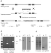
Figure 1
Figure 1
Targeted disruption of exon 3 of the Rs1h gene. (a) In the targeting construct, 1 bp of exon 3 (E3*), all of intron 3, and 66 bp of exon 4 (E4*) are deleted and replaced with a lacZ-neor cassette. (b) PCR amplification demonstrates the correct targeting of Rs1h. The relative positions of oligonucleotide primers and expected product sizes are given in a. (c) By using a probe spanning exons 4 to 6 of Rs1h, Northern blot analyses reveal the expected 5.6- and 4.9-kb transcripts in eye total RNA from the wt (wt) but not from the Rs1h knockout (−/Y) male mouse. Subsequent hybridization with a lacZ probe exhibits a fusion transcript of 3.7 kb only in the Rs1h-deficient animal. (d) Western blot analysis using eye cup protein extracts. Polyclonal antibody pAB-ap3RS1 labels the 24-kDa RS1 protein in WT mice. With the same antibody, the expected fusion protein of 120 kDa is not observed in mutant males, although it contains the antibody epitope, thus indicating that the targeted allele represents a true null allele. Equal loading of protein extracts is demonstrated by Coomassie staining.
Figure 2
Figure 2
Macromorphological evaluation of the Rs1h−/Y retina with scanning laser ophthalmoscopy. (a) Survey of the fundus, demonstrating a layer of cyst-like elevations in the inner retina. (b) Optical magnification reveals that the densely packed structures are clearly demarcated from the surrounding normal-appearing regions. (c) Focus on the retinal surface shows superficial vessels and the nerve fiber layer. Visible in the lower right quadrant are several larger cysts, one displacing a retinal vessel (arrow). (d) Fundus photograph of a patient with RS, featuring typical small macular cysts arranged in a stellate pattern (arrow) and radial striae centered on the fovea. There is an obvious similarity to the appearance of the mouse retina as shown in b.
Figure 3
Figure 3
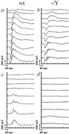
Electrophysiology of Rs1h−/Y and wt mice. Scotopic intensity series of a wt (a) and an Rs1h mutant mouse (b). Log light intensities (from top to bottom) were −4, −3, −2, −1.5, −1, −0.5, 0, 0.5, 1, 1.5 log cd⋅s/m2. The overall loss of amplitude and the additional selective reduction of the b-wave are clearly visible. Photopic intensity series of a wt control (c) and an Rs1h-deficient mouse (d). Log light intensities (from top to bottom) were −2, −1.5, −1, −0.5, 0, 0.5, 1, 1.5 log cd⋅s/m2. The photopic ERG of the Rs1h−/Y mice is strongly reduced, indicating a much more severe cone than rod dysfunction.
Figure 4
Figure 4
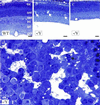
(ad) Semithin retinal sections of wt (WT) and Rs1h−/Y (−/Y) mice at 2 months of age. In meridional sections, the thickness of the central retina is markedly reduced in Rs1h−/Y mice (b and c) as compared with wt mice (a). In some Rs1h−/Y eyes, photoreceptor outer segments (POS) are present (b), whereas others show partial or complete absence of the POS (c). In Rs1h−/Y eyes with areas of preserved POS, large gaps are present between the cells of the INL (arrows in b). Such gaps are absent in areas with complete degeneration of POS (c). (d) Oblique tangential section through the INL of an Rs1h−/Y retina reveals large extracellular gaps (white arrows). In some of the gaps, cell bodies of microglia are observed (black arrow). os, photoreceptor outer segments; onl, outer nuclear layer; opl, outer plexiform layer, inl, inner nuclear layer; ipl, inner plexiform layer; gcl, ganglion cell layer. [Bars = 25 μm (ac) and 2.8 μm (d).]
Figure 5
Figure 5
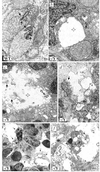
(af) Electron microscopy of the retina of wt (WT) and Rs1h−/Y (−/Y) mice at 2 months of age. (a) In the retina of wt mice, typical ribbon synapses are present at the photoreceptor terminals (black arrows). (b) In the retina of Rs1h−/Y mice, increased extracellular spaces (open arrow) are observed in regions of ribbon synapses (solid arrow). Larger extracellular gaps are present between individual photoreceptor terminals (asterisk). (c) The extracellular gaps (asterisk) in the INL of Rs1h−/Y mice are filled with cellular debris (solid arrows) and membranous whorls (open arrow). (d) Part of the extracellular debris in the INL gaps (asterisk) of Rs1h−/Y mice consists of fragmented nerve cell terminals (solid arrow) containing synaptic vesicles. (e) Cells with ultrastructural characteristics of microglia in the retina of Rs1h−/Y mice. In the increased extracellular spaces, cells with long cytoplasmic processes (arrows) are observed. (f) Upon higher magnification, multiple clear vesicles (solid arrow) and electron-dense phagolysosomes (open arrow) are observed in the cytoplasm of the cells. [Bars = 0.53 μm (a, b, d, and f) and 1.4 μm (c and e).]
Figure 6
Figure 6
Immunofluorescence microscopy of retinal cryosections from 2-month-old Rs1h−/Y and wt mice. (a and b) Rs1 labeling with the Rs1 3R10 monoclonal antibody (red). Image is merged with DAPI nuclear staining (blue) and differential interference contrast (DIC) microscopy. (c and d) Rhodopsin staining with the Rho 1D4 monoclonal antibody. (e and f) Cone opsin labeling with a mixture of polyclonal antibody JH 455 and JH 492. (Insets) Bar = 10 μm. (g and h) PAN-SAP antibody labeling of PSD-95 in the OPL and IPL in the wt mouse compared with the IS and OPL in the Rs1h−/Y mouse. Image is merged with DIC image showing the retinal layers. (i and j) Labeling of bipolar cells with the monoclonal antibody Mab 115A10. (k and l) Labeling of Mueller cells and retinal pigment epithelial (RPE) cells with an anti-CRALBP antibody. Abbreviations used are as in Fig. 4, plus (is), inner segment.

References

    1. Kellner U, Brummer S, Foerster M H, Wessing A. Arch Clin Exp Ophthalmol. 1990;228:432–437. - PubMed
    1. George N D, Yates J R, Moore A T. Br J Ophthalmol. 1995;79:697–702. - PMC - PubMed
    1. Puech B, Kostrubiec B, Hache J C, Francois P. J Fr Ophtalmol. 1991;4:153–164. - PubMed
    1. Yanoff M, Kertesz-Rahn E, Zimmerman L E. Arch Ophthalmol. 1968;79:49–53. - PubMed
    1. Manschot W A. Arch Ophthalmol. 1972;88:131–138. - PubMed

Publication types