

Immunological and pathological comparative analysis between experimental latent tuberculous infection and progressive pulmonary tuberculosis
- PMID: 11985512
- PMCID: PMC1906395
- DOI: 10.1046/j.1365-2249.2002.01832.x
Immunological and pathological comparative analysis between experimental latent tuberculous infection and progressive pulmonary tuberculosis
Abstract
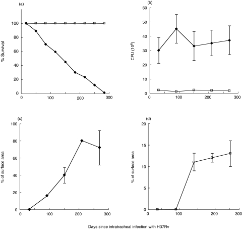
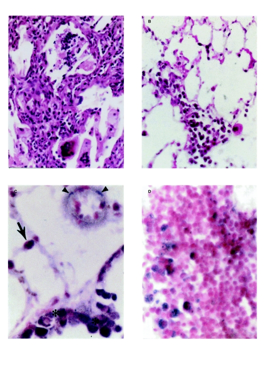
Mycobacterium tuberculosis produces latent infection or progressive disease. Indeed, latent infection is more common since it occurs in one-third of the world's population. We showed previously, using human material with latent tuberculosis, that mycobacterial DNA can be detected by in situ PCR in a variety of cell types in histologically-normal lung. We therefore sought to establish an experimental model in which this phenomenon could be studied in detail. We report here the establishment of such a model in C57Bl/6 x DBA/2 F1 hybrid mice by the intratracheal injection of low numbers of virulent mycobacteria (4000). Latent infection was characterized by low and stable bacillary counts without death of animals. Histological and immunological study showed granulomas and small patches of alveolitis, with high expression of tumour necrosis factor alpha (TNFalpha), inducible nitiric oxide synthase (iNOS), interleukin 2 (IL-2) and interferon gamma (IFNgamma). In contrast, the intratracheal instillation of high numbers of bacteria (1 x 10⁶) produced progressive disease. These animals started to die after 2 months of infection, with very high bacillary loads, massive pneumonia, falling expression of TNF-alpha and iNOS, and a mixed Th1/Th2 cytokine pattern. In situ PCR to detect mycobacterial DNA revealed that the most common positive cells in latently-infected mice were alveolar and interstitial macrophages located in tuberculous lesions, but, as in latently-infected human lung, positive signals were also seen in bronchial epithelium, endothelial cells and fibroblasts from histologically-normal areas. Our results suggest that latent tuberculosis is induced and maintained by a type 1 cytokine pattern plus TNFalpha, and that mycobacteria persist intracellularly in lung tissue with and without histological evidence of a local immune response.
Figures
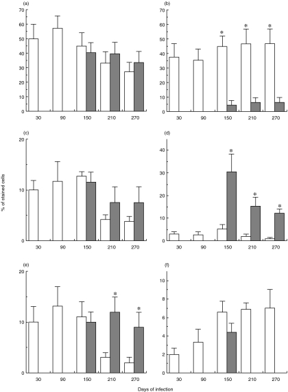
 ) and latent infection (□). Pooled results from two experiments, each with eight mice, are expressed as the mean and standard deviation. Asterisks indicate statistical significance (P < 0·005). (a) IL-2; (b) IL-4; (c) TNF; (d) IL-1; (e) iNOS; (f) BCG.
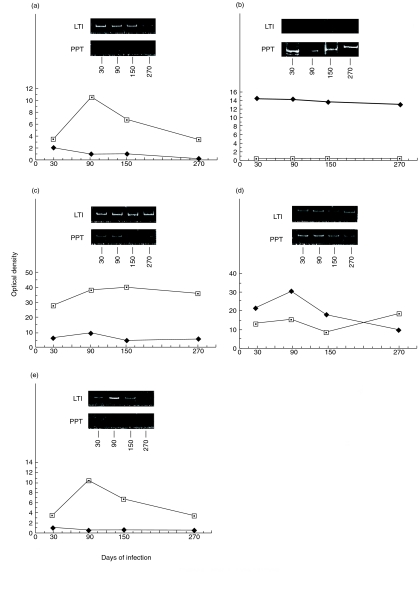
References
-
- Parrish NM, Dick JD, Bishai WR. Mechanisms of latency in Mycobacterium tuberculosis. Trends Microbiol. 1998;6:107–12. - PubMed
-
- Kochi A. The global tuberculosis situation and the new control strategy of the World Health Organization. Tubercle. 1991;72:1–6. - PubMed
-
- Wayne LG. Dormancy of Mycobacterium tuberculosis and latency of disease. Eur J Clin Microbiol Infect Dis. 1994;13:908–14. - PubMed
Publication types
MeSH terms
Substances
LinkOut - more resources
Full Text Sources
Other Literature Sources
