Review
. 2002 Apr;105(4):375-90.
doi: 10.1046/j.1365-2567.2002.01375.x.

Tissue-specific expression of mast cell granule serine proteinases and their role in inflammation in the lung and gut

Affiliations
Review

Tissue-specific expression of mast cell granule serine proteinases and their role in inflammation in the lung and gut

Hugh R P Miller et al. Immunology. 2002 Apr.

Abstract

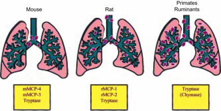
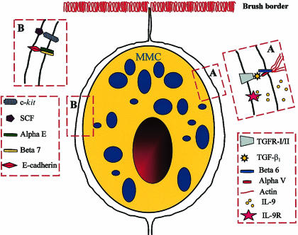
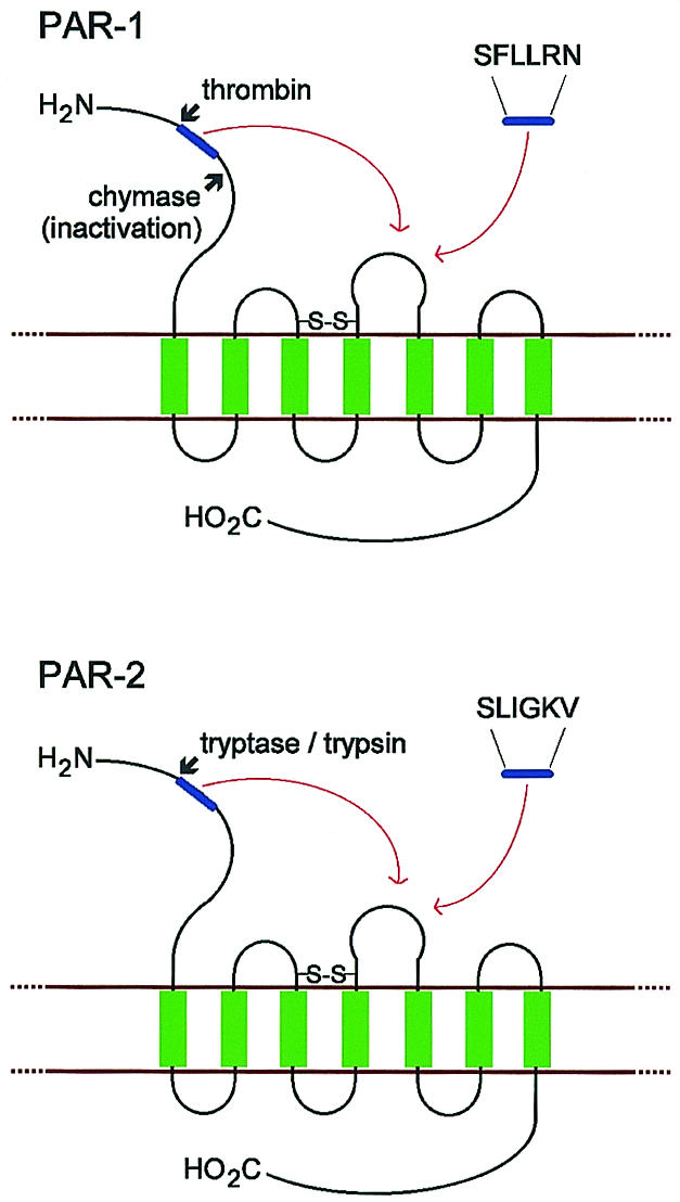
Serine proteinases with trypsin-like (tryptase) and chymotrypsin-like (chymase) properties are major constituents of mast cell granules. Several tetrameric tryptases with differing specificities have been characterized in humans, but only a single chymase. In other species there are larger families of chymases with distinct and narrow proteolytic specificities. Expression of chymases and tryptases varies between tissues. Human pulmonary and gastrointestinal mast cells express chymase at lower levels than tryptase, whereas rodent and ruminant gastrointestinal mast cells express uniquely mucosa-specific chymases. Local and systemic release of chymases and tryptases can be quantified by immunoassay, providing highly specific markers of mast cell activation. The expression and constitutive extracellular secretion of the mucosa-specific chymase, mouse mast cell proteinase-1 (mMCP-1), is regulated by transforming growth factor-beta1 (TGF-beta1) in vitro, but it is not clear how the differential expression of chymases and tryptases is regulated in other species. Few native inhibitors have been identified for tryptases but the tetramers dissociate into inactive subunits in the absence of heparin. Chymases are variably inhibited by plasma proteinase inhibitors and by secretory leucocyte protease inhibitor (SLPI) that is expressed in the airways. Tryptases and chymases promote vascular permeability via indirect and possibly direct mechanisms. They contribute to tissue remodelling through selective proteolysis of matrix proteins and through activation of proteinase-activated receptors and of matrix metalloproteinases. Chymase may modulate vascular tissues through its ability to process angiotensin-I to angiotensin-II. Mucosa-specific chymases promote epithelial permeability and are involved in the immune expulsion of intestinal nematodes. Importantly, granule proteinases released extracellularly contribute to the recruitment of inflammatory cells and may thus be involved in innate responses to infection.


Figures

Figure 1
Figure 1
Schematic representation of the distribution of mast cells in the lungs of mouse, rat, primates and ruminants. Note that the larger mammals have a substantial proportion of mast cells in the lung parenchyma, whereas in the mouse, the mast cells are located predominantly adjacent to the major airways. The proteinases that are predominantly expressed in the airways are shown in the boxes below each diagram. mMCP, mouse mast cell proteinase; rMCP, rat mast cell proteinase.
Figure 2
Figure 2
Confocal image (a) of 14-day-old-mouse bone marrow cultures demonstrating the presence of mature mast cells with abundant granules containing mouse mast cell proteinase-1 (green fluorescence) and expressing the integrin αE (red fluorescence) on their surface membranes. A light micrograph (b) of the mouse bone marrow mast cells (mBMMC) stained with Leishman's shows that they are mature, heavily granulated cells. The cells were grown in medium containing recombinant mouse interleukin (IL)-3, IL-9 and stem cell factor (SCF) supplemented with recombinant human transforming growth factor-β1 (TGF-β1), as described in detail by Miller et al. (Horizontal bars represent 10 µm.)
Figure 3
Figure 3
Diagrammatic representation of a mouse mucosal mast cell (mMMC) within the intestinal epithelium with the postulated receptor–ligand interactions between the two cell types illustrated in boxes A and B. In Box A the epithelial cell-specific integrins αVβ6 are shown binding an activated transforming growth factor-β1 (TGF-β1) molecule and presenting it to its receptor on the mast cell surface. The probable interaction between the integrins and actin fibres in the epithelial cytoskeleton is also illustrated. We speculate that interleukin (IL)-9 is produced by the epithelium, as indicated by published studies, and that it binds to its receptor on the mast cell surface. In box B the interaction between epithelially expressed stem cell factor (SCF) and its tyrosine kinase receptor c-kit is shown together with the probable interaction between the integrins αEβ7 on the mast cell surface and epithelially expressed E-cadherin. The receptor–ligand interactions illustrated here are consistent with in vitro studies showing that IL-9, SCF and TGF-β1 are important growth and differentiation factors. We speculate that the constitutive secretion of mouse mast cell proteinase-1 (mMCP-1), induced by TGF-β1, exerts a modulatory effect on these receptor–ligand interactions through, for example, the proteolytic degradation of ligands such as SCF or of the cytokines in the intercellular milieu. This hypothesis might explain the augmented mast cell hyperplasia in mMCP-1−/− mice lacking this proteinase.,
Figure 4
Figure 4
Schematic representation of human proteinase-activated receptor-1 (PAR-1) and PAR-2. The N-terminus is extracellular, the C-terminus is intracellular and transmembrane regions are shown in green. Activation of PAR-1 by thrombin and PAR-2 by tryptase or trypsin exposes the tethered ligand region (shown in blue). This docks into the binding region of extracellular loop 2, which can also be activated by a synthetic hexapeptide representing the new N-terminus. The inactivation of PAR-1 by chymase is also represented, which is presumed to be via cleavage C-terminal to the tethered ligand region.

References

    1. Bairoch A, Apweiler R. The SWISS-PROT protein sequence database and its supplement TrEMBL in 2000. Nucl Acids Res. 2000;28:45–8. - PMC - PubMed
    1. Miller HR. Mucosal mast cells and the allergic response against nematode parasites. Vet Immunol Immunopathol. 1996;54:331–6. - PubMed
    1. Enerback L. Mucosal mast cells in the rat and in man. Int Arch Allergy Appl Immunol. 1987;82:249–55. - PubMed
    1. Befus D. Intestinal mast cell polymorphism: new research directions and clinical implications. J Pediatr Gastroenterol Nutr. 1986;5:517–21. - PubMed
    1. Irani AM, Schwartz LB. Human mast cell heterogeneity. Allergy Proc. 1994;15:303–8. - PubMed

Publication types

LinkOut - more resources