. 2002 May;9(3):677-86.
doi: 10.1128/cdli.9.3.677-686.2002.

Low-dose UVB contributes to host resistance against Leishmania amazonensis infection in mice through induction of gamma interferon and tumor necrosis factor alpha cytokines

Affiliations

Low-dose UVB contributes to host resistance against Leishmania amazonensis infection in mice through induction of gamma interferon and tumor necrosis factor alpha cytokines

Noor Mohammad Khaskhely et al. Clin Diagn Lab Immunol. 2002 May.

Abstract

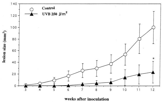
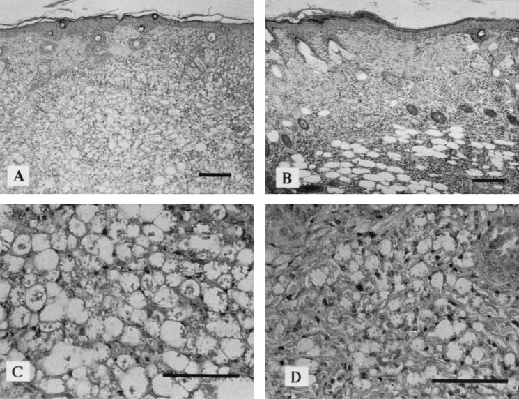
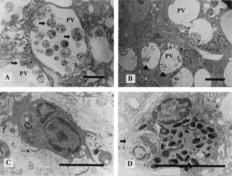
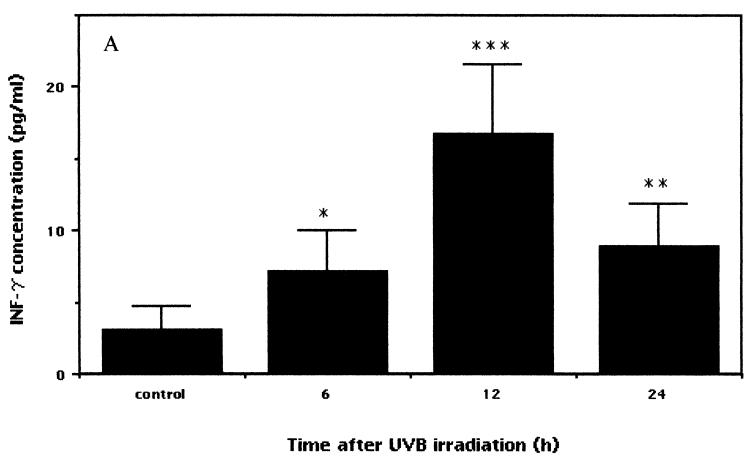
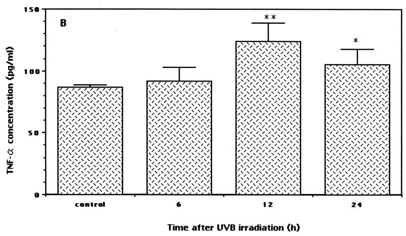
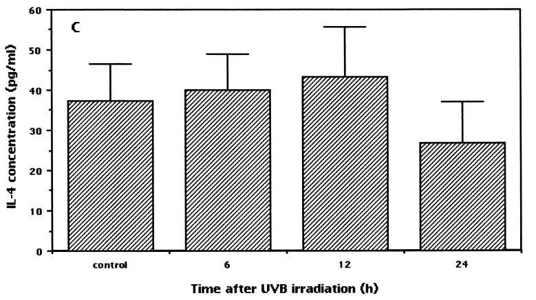
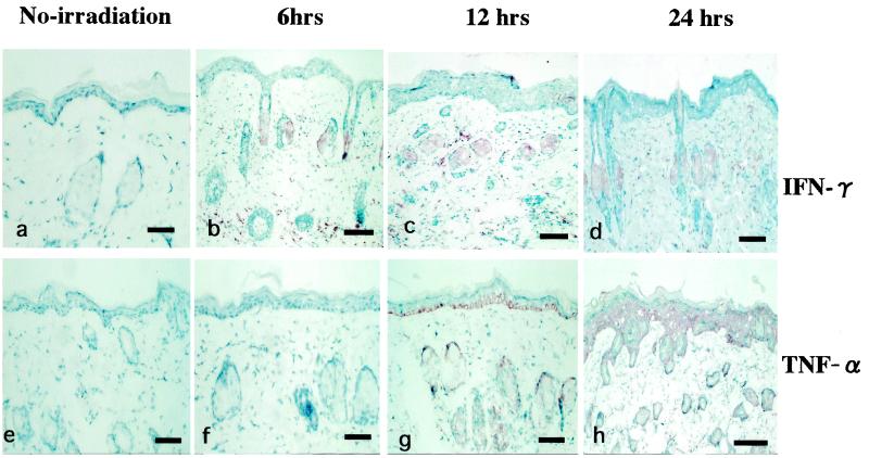
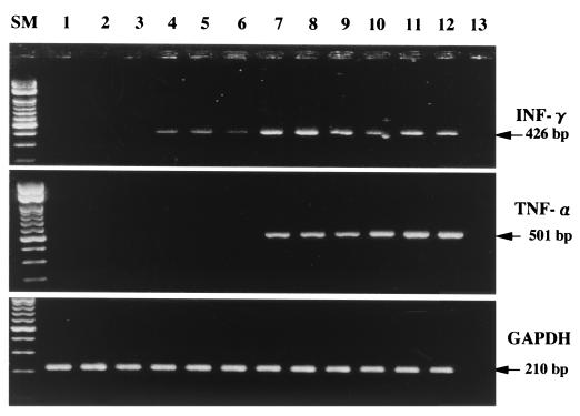
UV radiation suppresses the immune response, a fact which raises the question of whether the phenomenon may find practical applications in the outcome of infectious diseases. In this study, BALB/c mice were exposed to low-dose UVB (250 J/m(2)) from Dermaray M-DMR-100 for 4 consecutive days. Twelve hours after the last UV exposure, groups of mice were injected with 2 x 10(6) Leishmania amazonensis promastigotes. The development of skin lesions, as assessed by measurement of visible cutaneous lesions, was significantly suppressed in low-dose UVB-irradiated mice compared to nonirradiated controls. In order to characterize the cytokines involved in this phenomenon, BALB/c mice were irradiated with identical doses of UVB, and gamma interferon (IFN-gamma), tumor necrosis factor alpha (TNF-alpha), and interleukin 4 cytokine levels in blood serum and skin were examined at different times by a sandwich enzyme-linked immunosorbent assay, immunohistochemical analysis, and reverse transcription (RT)-PCR. Upregulated expression of serum IFN-gamma and TNF-alpha was observed from 6 to 24 h. Positive results for IFN-gamma and TNF-alpha in UVB-irradiated mice were obtained by immunohistochemical analysis. By RT-PCR, the mRNA expression of both IFN-gamma and TNF-alpha cytokines was detected in a time-dependent manner only in UVB-irradiated mice. Histopathological analysis and electron microscopy revealed that cellular infiltration, tissue parasitism, and parasitophorus vacuoles in irradiated mice were markedly less noticeable than those in nonirradiated controls. These results suggested that low-dose UVB irradiation played a pathogen-suppressing role in Leishmania-susceptible BALB/c mice via systemic and local upregulation of Th1 (IFN-gamma and TNF-alpha) cytokines.


Figures

FIG. 1.
FIG. 1.
Low-dose UVB irradiation suppresses the lesion development of cutaneous L. amazonensis infection. BALB/c mice were irradiated with UVB at 250 J/m2 for 4 consecutive days; 12 h later, control and irradiated mice were injected with 2 × 10⁶ L. amazonensis promastigotes. Three weeks after inoculation, visible skin lesions were measured weekly, and size was calculated by measuring length and width. Nonirradiated and irradiated mice were killed at 12 weeks after inoculation. Data presented here are the means and standard deviations from a representative experiment repeated three times with the same results. Statistically, the significance of the difference between the groups was analyzed by Student's unpaired t test. An asterisk indicates a P value of <0.0008.
FIG. 2.
FIG. 2.
Low-dose UVB irradiation decreases parasite burden at the parasite injection site. Hematoxylin-eosin-stained tissue specimens were from BALB/c mice injected with 2 × 10⁶ L. amazonensis promastigotes. (A and C) Tissue samples taken from control mice. (B and D) Tissue samples taken from mice irradiated with low-dose UVB. Bars, 100 μm.
FIG. 3.
FIG. 3.
Transmission electron micrographs of parasite inoculation sites after 12 weeks. (A) A specimen from a control mouse shows large PV, containing many Leishmania parasites (arrows). (B) A specimen from a low-dose UVB-irradiated mouse shows small PV, containing few Leishmania parasites; some show degenerative changes (arrowheads). (C) In a low-dose UVB-irradiated mouse, signs of macrophage activation, a large number of lysosomes, and many microvilli (arrows) are seen. (D) Leishmania parasites are phagocytized by eosinophils in a low-dose UVB-irradiated mouse (arrow). Scale bars, 5 μm.
FIG. 4.
FIG. 4.
Expression of serum cytokine levels in low-dose UVB-irradiated and control mice. After the last exposure to low-dose irradiation, blood was obtained at various times. Serum samples obtained by centrifugation at 2,000 × g for 20 min at 4°C were analyzed with commercially available ELISA kits to measure IFN-γ, TNF-α, and IL-4 protein concentrations. The data shown represent the time-dependent induction of IFN-γ (A) (asterisk, P < 0.02; double asterisks, P < 0.004; and triple asterisks, P < 0.0003), TNF-α (B) (asterisk, P < 0.03; and double asterisks, P < 0.005), and IL-4 (C) after low-dose UVB irradiation. Each sample was analyzed in duplicate, and data are expressed as means and standard deviations from a representative experiment repeated three times with the same results. Statistically, the significance of the difference between the groups was analyzed by Student's unpaired t test.
FIG. 4.
FIG. 4.
Expression of serum cytokine levels in low-dose UVB-irradiated and control mice. After the last exposure to low-dose irradiation, blood was obtained at various times. Serum samples obtained by centrifugation at 2,000 × g for 20 min at 4°C were analyzed with commercially available ELISA kits to measure IFN-γ, TNF-α, and IL-4 protein concentrations. The data shown represent the time-dependent induction of IFN-γ (A) (asterisk, P < 0.02; double asterisks, P < 0.004; and triple asterisks, P < 0.0003), TNF-α (B) (asterisk, P < 0.03; and double asterisks, P < 0.005), and IL-4 (C) after low-dose UVB irradiation. Each sample was analyzed in duplicate, and data are expressed as means and standard deviations from a representative experiment repeated three times with the same results. Statistically, the significance of the difference between the groups was analyzed by Student's unpaired t test.
FIG. 5.
FIG. 5.
Immunohistochemical detection of IFN-γ and TNF-α cytokine expression at different times after low-dose UVB irradiation. (a and e) Samples from nonirradiated mice used as controls. (b and f, c and g, and d and h) Samples from mice at 6, 12, and 24 h after the last UVB exposure, respectively.
FIG. 6.
FIG. 6.
Detection by RT-PCR assays of IFN-γ, TNF-α, and GAPDH mRNAs in tissues from nonirradiated control mice and low-dose UVB-irradiated mice at different times. A total of six mice per group were used. Results for representative samples taken from three mice in each group are shown. Lanes 1 to 3, nonirradiated mice; lanes 4 to 6, 7 to 9, and 10 to 12, mice at 6, 12, and 24 h after the last UVB irradiation, respectively. Lane 13, no DNA added (negative control). Lane SM, size markers.

References

    1. Afonso, L. C., and P. Scott. 1993. Immune responses associated with susceptibility of C57BL/10 mice to Leishmanaia amazonensis. Infect. Immun. 61:2952-2959. - PMC - PubMed
    1. Almeida, R. P., M. Barral-Netto, A. Ribeiro de Jesus, L. A. R. Freitas, E. M. Carvalho, and A. Barral. 1996. Biological behavior of Leishmania amazonensis isolated from humans with cutaneous, mucosal or visceral leishmaniasis in BALB/c mice. Am. J. Trop. Med. Hyg. 54:178-184. - PubMed
    1. Andrade, Z. A., S. G. Reed, S. Roters, and M. Sadigursky. 1984. Immuno-patology of experimental cutaneous leishmaniasis. Am. J. Pathol. 114:137-148. - PMC - PubMed
    1. Araneo, B. A., T. Dowell, H. B. Moon, and R. A. Daynes. 1989. Regulation of murine lymphokine production in vivo: ultraviolet radiation exposure depresses IL-2 and enhances IL-4 production by T cells through an IL-1-dependent mechanism. J. Immunol. 143:1737-1744. - PubMed
    1. Barrel-Netto, M., J. S. daSilva, A. Barrel, and S. Reed. 1995. Up-regulation of T helper 2 and down-regulation of T helper 1 cytokines durin murine retrovirus-induced immunodeficiency syndrome enhances susceptibility of a resistant mouse strain to Leishmania amazonensis. Am. J. Pathol. 146:635-642. - PMC - PubMed

Publication types

MeSH terms

LinkOut - more resources